
Animal Tissues
Think of an animal’s body as a sophisticated building. Every building needs a skin (covering), a skeleton and muscle (support and movement), a communication network (nerves), and a fluid transport system (blood). Animal tissues are precisely these four systems. In this section, we will concentrate on the outermost one — the covering tissue.
Epithelial Tissue
The word “epithelium” comes from the Greek epi (upon) + thele (nipple) — historically, upon a surface. Think of epithelial tissue as the body’s wallpaper — it lines every surface, every cavity, every tube, every organ. It is the first thing the outside world touches, and the last line of defence before sensitive internal organs.
Three fundamental characteristics define all epithelial tissue: cells are tightly packed (no room for pathogens to sneak in), separated from deeper tissues by a basement membrane (a fibrous scaffold that anchors and nourishes), and their permeability is tuned — some let things through easily (like lung alveoli for gas exchange), others are near-impermeable fortresses (like skin).
Now here is the elegant logic of the entire classification: epithelial cells are named in two ways — by how many layers they form (simple = one layer, stratified = many), and by the shape of the cells (squamous = flat, cuboidal = cube, columnar = tall column). Once you know these two axes, the entire chart maps itself.
Simple Epithelium
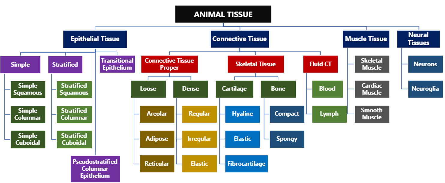
A single layer of cells lining a surface sounds fragile, but it is actually perfectly engineered for efficiency. When things need to pass through quickly — oxygen, nutrients, waste — a single thin layer is ideal. Nature puts exactly as much barrier as it needs, and not a cell more.
Simple squamous epithelium — the flattest, thinnest cells of all. Imagine floor tiles — flat and seamless. Their thinness is their superpower: substances diffuse across them with ease. This is precisely why they line the alveoli (air sacs of the lungs, where oxygen must rapidly enter the blood) and the endothelium of blood vessels (where nutrients and gases constantly cross).
Also note: when simple squamous epithelium lines blood and lymph vessels specifically, it earns the special name endothelium; when it lines the body’s inner cavities (pleura, pericardium), it is called mesothelium.
Simple cuboidal epithelium — cells that look like dice from the side. Equal height and width. Designed for secretion and absorption, not just passive diffusion. Found in kidney tubules (which actively reabsorb glucose, salts, and water from filtered blood), glandular ducts, and the thyroid gland (which secretes hormones). The cubic shape indicates a busy, metabolically active cell — more volume, more machinery.
Simple columnar epithelium — tall, upright cells that look like columns in a Greek temple. The free surface (the exposed top) may have microvilli — tiny finger-like projections that massively increase the absorptive surface area. This tissue lines the digestive tract (gut), uterine tubes, and gall bladder. The columnar shape signals a heavy-duty absorption and secretion role — more height = more space for the secretory/absorptive machinery.
Two important variants deserve special mention here.
- Ciliated epithelium uses columnar or cuboidal cells topped with cilia — tiny hair-like projections that beat rhythmically to sweep particles or mucus in a specific direction. Found in the bronchioles (sweeping dust and pathogens out of the lungs) and the fallopian tubes (propelling the egg towards the uterus).
- Glandular epithelium consists of specialised cells built for secretion — either unicellular (like goblet cells that produce mucus in the alimentary canal) or multicellular (like salivary glands).
This connects to the critical distinction between Exocrine glands (which release their products through a duct — saliva, sweat, mucus, digestive enzymes) and Endocrine glands (which have no duct and pour hormones directly into the surrounding fluid and bloodstream). “Exo” means outside, “endo” means inside — the duct is the deciding criterion.
Stratified Epithelium — Protection Over Efficiency
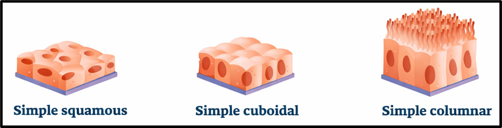
Where a single layer is enough for exchange, multiple layers are needed for protection. Wherever the body faces friction, abrasion, or mechanical stress, stratified epithelium stands guard. The trade-off is clear: multiple layers block rapid exchange, but they can absorb punishment that would destroy a single layer instantly.
Stratified squamous epithelium is the king of protection. Multiple layers of flat cells — the outermost ones often dying and becoming keratin-filled shields (as in the skin’s epidermis). Found in the skin’s outer layer, oral cavity, oesophagus, and vagina — all surfaces that experience constant friction. The reason your skin can take a scraped knee without exposing internal tissue is entirely due to this tissue.
Stratified cuboidal epithelium — multiple layers of cube-shaped cells, offering protection while retaining some secretory ability. Found in sweat glands, mammary glands, and salivary glands.
Stratified columnar epithelium — multiple layers of tall cells, found in the male urethra and certain gland ducts — again, balancing protection with secretion.
Two Special Cases
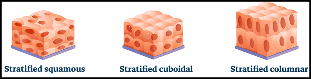
Pseudostratified columnar epithelium — this is a masterclass in biology’s capacity to deceive. It looks like multiple layers because the cells have nuclei at different heights, creating the optical illusion of strata. But pull back and look carefully: every single cell touches the basement membrane. It is — by the strictest definition — a single layer. “Pseudo” means false.
It is ciliated and secretes mucus. Found precisely where you’d expect such a tissue: the respiratory tract (trachea and bronchi), where cilia sweep mucus and trapped particles upward away from the lungs, and parts of the male reproductive system.
Transitional epithelium — the shapeshifter. Found in the urinary bladder, ureters, and part of the urethra — organs that must dramatically expand and contract. When the bladder is empty, cells are domed and piled in thick layers. When the bladder fills and stretches, those same cells flatten out and slide past one another. The tissue transitions between states. No other epithelium does this — transitional epithelium is uniquely designed for organs that change volume.
Cell Junctions — The Invisible Infrastructure
Here is a concept that often gets skimmed in textbooks but is fundamental to understanding how epithelial tissue actually works as a barrier and communication system.
Individual cells would be useless without connections to their neighbours. Three types of junctions hold the cellular community together:
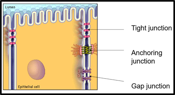
Tight junctions are the body’s leak-proof seals. They fuse adjacent cell membranes so tightly that nothing can pass between the cells — everything must go through the cell itself, allowing the cell to act as a selective gatekeeper. The blood-brain barrier is largely maintained by tight junctions, which is why only very specific molecules can enter the brain.
Adhering junctions are the biological cement. They use protein filaments (cadherin molecules attached to the cytoskeleton) to rivet cells together and give tissue mechanical integrity — the ability to withstand stretching and shearing forces. Think of them as the stitching that holds a fabric together. Critical in heart muscle, which is under constant mechanical stress.
Gap junctions are the most sophisticated of the three — they are not about sealing or sticking, but about talking. These are aqueous channels (made of proteins called connexins) that physically connect the cytoplasm of two adjacent cells, allowing ions, small molecules, and electrical signals to pass directly from one cell to another. This is what allows the heart to beat in synchrony — one cell’s electrical impulse passes instantly to its neighbour through gap junctions, creating a coordinated wave.
The Thread That Connects Everything
Notice the consistent biological logic: wherever the demand is exchange (gas, nutrients), you get a single thin layer — simple squamous. Wherever the demand is active transport (secretion, absorption), you get a taller, more metabolically active cell — cuboidal or columnar. Wherever the demand is protection against friction, you get multiple layers — stratified. And whenever an organ must change shape, you get a tissue that can stretch — transitional.
Let’s move ahead now!
Connective Tissues
If epithelial tissue was the covering, connective tissue is the skeleton of everything else — the glue, the scaffold, the highway, and the armour of the body, all rolled into one tissue type.
Here is the single most important insight to hold before we begin: connective tissue is defined by what is between the cells, not the cells themselves. In every other tissue, the cells are the story. In connective tissue, the extracellular matrix (ECM) is the story. The cells are merely the workers who build and maintain it.
The Extracellular Matrix
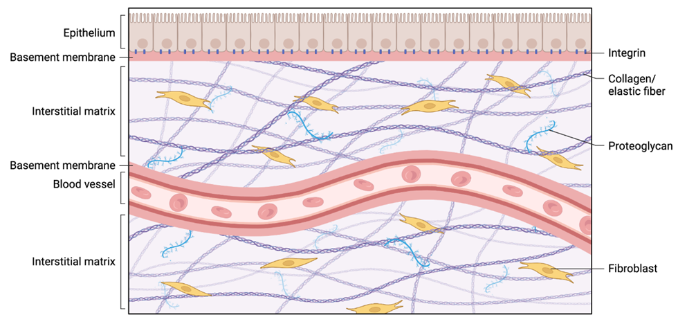
The ECM is produced and maintained by fibroblast cells. It has two components.
- First, protein fibres — collagen (which gives tensile strength, like steel cables in concrete) and elastin (which gives the ability to stretch and snap back, like a rubber band).
- Second, ground substance — a gel-like filler made of proteoglycans, glycosaminoglycans (GAGs), and glycoproteins that cushions cells, facilitates nutrient-waste exchange, and holds the whole structure together like mortar between bricks.
The genius is in the variation: change the ECM composition, and you get a completely different tissue — fluid blood, flexible cartilage, rigid bone. Same principle, radically different outcomes.
Connective Tissue Proper
Loose Connective Tissue
Think of loose connective tissue as bubble wrap — loosely arranged fibres and cells with plenty of space in between.
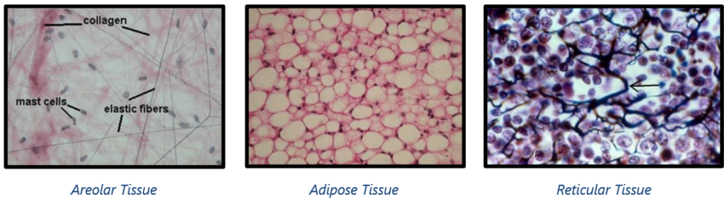
Areolar tissue is the most widespread tissue in the body. It is a loose arrangement of collagen and elastin fibres in a gel-like ground substance, filled with various cell types including fibroblasts and mast cells.
- It fills the spaces inside organs, holds organs in their positions, stores water and salts, and is the first responder in tissue repair. Found under the skin and wrapped around blood vessels, nerves, and organs — essentially, the universal packing material of the body.
- If you have ever seen the loose, wispy tissue that connects a chicken’s skin to its muscle, that is areolar tissue.
Adipose tissue is specialised loose connective tissue where fat cells called adipocytes dominate and the ECM is minimal.
- Each adipocyte is essentially a balloon filled with a lipid droplet, with the nucleus pushed to one side. Its three jobs are energy storage (the body’s long-term fuel tank), thermal insulation (keeping core temperature stable), and mechanical cushioning (protecting kidneys, eyeballs, and other organs from impact).
- Located under the skin as subcutaneous fat, around internal organs as visceral fat, and in bone marrow.
Reticular tissue forms a delicate, mesh-like network of reticular fibres (a fine type of collagen). Think of it as a biological sponge or fishing net.
- It provides the structural scaffold for soft organs that need to hold many cells in an organised but flexible arrangement — the liver, spleen, lymph nodes, and bone marrow all depend on reticular tissue as their internal framework. It also helps filter blood and lymph as they pass through these organs.
Dense Connective Tissue
Where loose connective tissue is bubble wrap, dense connective tissue is rope. The defining feature is a high density of collagen fibres, leaving very little space for cells or ground substance.
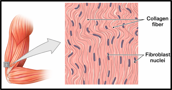
Dense regular connective tissue has collagen fibres arranged in neat, parallel bundles — like a bundle of cables all running the same direction.
- This gives extraordinary strength in one direction. This is the tissue of tendons (muscle to bone) and ligaments (bone to bone).
- When you strain a tendon or ligament, this is the tissue being overstretched or torn. The parallel arrangement is the key point — unidirectional tensile strength.
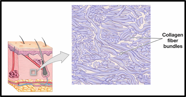
Dense irregular connective tissue has the same density of collagen, but the fibres are arranged in a random, criss-cross pattern — strength in all directions simultaneously. Found in the dermis (deep layer of skin), the capsules that envelop organs like the kidney and liver, and the periosteum (the membrane covering bones). The irregular arrangement makes perfect sense: the skin, for instance, must resist pulling forces from any direction.
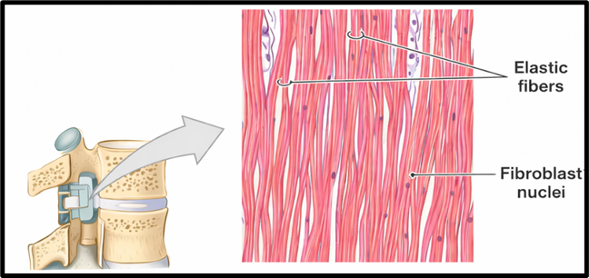
Elastic tissue contains a high density of elastin fibres — it can stretch significantly and then snap back to its original length. Found in the walls of large arteries (the aorta must expand with every heartbeat and recoil to push blood forward), bronchial tubes, and certain ligaments like the ligamentum flavum of the spine. This is why arteries are described as “elastic” — they literally are.
Skeletal Tissue
Cartilage
Cartilage is the elegant middle ground between soft tissue and bone. Its cells are called chondrocytes, which sit in small cavities called lacunae within a matrix of collagen and proteoglycans.
The critical point: cartilage is avascular (no blood vessels) and aneural (no nerves). This explains why cartilage damage heals so slowly — with no blood supply, there is no easy delivery of repair materials.
Three types, distinguished by what is in their matrix:
Hyaline cartilage — “hyaline” means glassy. It is the most common and the template cartilage of the embryo (most bones begin as hyaline cartilage and are later replaced by bone in a process called ossification). Found at the ends of long bones (articular cartilage), in the nose, trachea, and ribs. Its glassy, smooth surface reduces friction between bones in joints.
Elastic cartilage — identical to hyaline except it has elastin fibres woven into the matrix, giving it the ability to bend and spring back. You can demonstrate this yourself: bend your ear. It springs back. That is elastic cartilage in action. Also found in the epiglottis — the flap that covers the larynx during swallowing.
Fibrocartilage — the toughest of the three. Thick bundles of collagen make it both strong and shock-absorbing. Precisely what you need between vertebrae (the intervertebral discs that absorb spinal compression) and in the knee joint meniscus. Notably, fibrocartilage is the tissue that forms in a healing bone fracture — a temporary bridge before true bone is laid down.
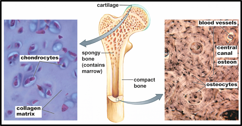
Bone
If cartilage is flexible scaffolding, bone is reinforced concrete. The matrix has two components working in concert: collagen fibres (providing elasticity — preventing the bone from shattering like chalk) and inorganic mineral salts, primarily calcium phosphate (providing hardness and compressive strength). Remove the minerals and bone becomes rubbery. Remove the collagen and bone becomes brittle. Both are essential.
Three cell types manage this living tissue:
- osteoblasts (the builders — they produce osteoid, the protein matrix that then mineralises into bone),
- osteocytes (mature osteoblasts now embedded in the matrix, sitting in spaces called lacunae, maintaining bone tissue), and
- osteoclasts (the demolition crew — they break down old bone during remodelling).
Bone is constantly being remodelled throughout life — osteoblasts and osteoclasts work in coordinated shifts.
Structurally, bone has two layers:
- compact (cortical) bone — the dense, solid outer shell that provides strength — and
- cancellous (spongy) bone — the inner honeycomb-like network containing bone marrow, where blood cells are produced.
Comparison between Bone and Cartilage
| Feature | Bone | Cartilage |
| Hardness | Hard and non-flexible | Flexible |
| Structure | Composed of calcium phosphate and collagen | Composed of collagen and proteoglycans |
| Blood Supply | Vascular (has blood vessels) | Avascular (lacks blood vessels) |
| Nerve Supply | Innervated (has nerves) | Aneural (lacks nerves) |
| Cavity | Present | Absent |
| Function | Support, protection, movement, blood cell production, mineral storage | Support, cushioning, flexibility |
Fluid Connective Tissue
Blood
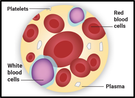
- Blood is connective tissue where the ECM is entirely liquid — the plasma. Plasma is 90% water carrying proteins, electrolytes, hormones, nutrients, and waste products.
- Suspended in plasma are the formed elements:
- erythrocytes (red blood cells — carry oxygen via haemoglobin, and transport CO₂ back),
- leukocytes (white blood cells — the immune army), and
- thrombocytes (platelets — fragments of large cells called megakaryocytes, essential for clotting).
Lymph
When blood passes through capillaries, some of the plasma seeps out into the spaces between cells, forming interstitial fluid or tissue fluid. Most of this is reabsorbed, but the remainder is collected by the lymphatic system and becomes lymph — a colourless fluid that flows back towards the heart through lymphatic vessels.
Components of Lymph
- Lymph Plasma
- Composed primarily of → Water (~95%), Electrolytes, Proteins (mainly albumin and globulins), Nutrients (glucose, lipids, etc.), Waste products and Various cells
- Comparison with blood plasma: Similar in composition
- Contains → Less calcium, Fewer blood proteins, Less phosphorus, Higher glucose concentration
- Lymph Corpuscles
- Mainly lymphocytes suspended in lymph plasma.
- Lymphocytes are white blood cells crucial for immune defense.
Types of Lymphocytes
i) T Cells
- Involved in cell-mediated immunity
- Recognize and destroy infected or cancerous cells
ii) B Cells
- Responsible for antibody (immunoglobulin) production
- Target specific antigens (foreign molecules)
iii) Natural Killer (NK) Cells
- Destroy infected or abnormal cells without prior exposure
- Part of innate immunity
Functions of Lymph
- Fluid balance: Returns excess interstitial fluid back to the bloodstream
- Immunity: Transports white blood cells to fight infections
- Fat absorption: Carries absorbed fats from intestines to the bloodstream
- Removal of waste products: Helps eliminate cellular waste
B Cells vs T Cells
| Feature | B Cells | T Cells |
| Primary function | Antibody production | Cellular immunity |
| Produced in | Bone marrow | Bone marrow |
| Mature in | Bone marrow | Thymus |
| Type of immunity | Humoral immunity | Cell-mediated immunity |
| Mechanism of action | Use antibodies to neutralise pathogens | Directly kill infected cells and regulate immune response |
Blood vs Lymph
| Feature | Blood | Lymph |
| Colour | Red | Colourless |
| Composition | Plasma, RBCs, WBCs, platelets | Plasma-like fluid, mainly WBCs |
| Function | Transport of O₂, CO₂, nutrients, waste, hormones; temperature regulation; immunity | Returns excess fluid, transports fats, immunity |
| Circulation | Through arteries, veins, capillaries | Through lymphatic vessels |
| Cells present | RBCs, WBCs, platelets | Mainly WBCs (lymphocytes) |
| Pressure | Higher | Lower |
| Flow rate | Faster | Slower |
| Direction | Closed circulatory system | One-way towards the heart |
Muscle Tissue
Muscular tissue is a mesodermal tissue specialized for movement. Whether it is walking, breathing, or even the beating of your heart—everything involves muscles.
At a fundamental level, muscles work because of four key properties:
- Excitability → ability to respond to stimuli
- Contractility → ability to shorten (generate force)
- Extensibility → ability to stretch
- Elasticity → ability to return to original shape
👉 This combination makes muscles dynamic rather than rigid structures.
Structure Insight
Each muscle is made up of:
- Long cylindrical muscle fibres
- These fibres contain myofibrils
- Myofibrils contain contractile proteins (mainly actin and myosin)
These proteins slide past each other → causing contraction and relaxation → resulting in movement
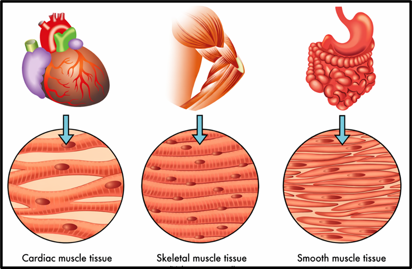
📊 Types of Muscle Tissue
Muscles are classified based on location and function:
| Type | Control | Location | Special Features |
| Skeletal | Voluntary | Attached to bones | Striated, multinucleated |
| Cardiac | Involuntary | Heart | Branched, intercalated discs |
| Smooth | Involuntary | Internal organs | Non-striated, spindle-shaped |
1️⃣ Skeletal Muscle
This is what you typically think of as “muscle”.
- Responsible for voluntary movements (walking, writing)
- Attached to bones via tendons
- Helps in posture and locomotion
Structural Features:
- Long, cylindrical fibres
- Multinucleated
- Striated appearance (due to actin & myosin arrangement)
👉 Think of it as a highly organized machine designed for precision movement.
2️⃣ Cardiac Muscle
Found only in the heart, this muscle ensures continuous blood circulation.
- Works involuntarily
- Contracts rhythmically to pump blood
Unique Features:
- Cells are short and branched
- Usually uninucleated
- Connected by intercalated discs
👉 Intercalated discs allow synchronised contraction, so the heart beats as a coordinated unit.
3️⃣ Smooth Muscle (Visceral Muscle)
These muscles are present in internal organs like → Alimentary canal, Blood vessels, Reproductive tract, Iris of the eye
Features:
- Spindle-shaped cells
- Uninucleated
- No striations → smooth appearance
Function:
- Involuntary movements like:
- Food movement (peristalsis)
- Movement of gametes
👉 These muscles ensure internal processes run smoothly without conscious effort.
Neural Tissue
Now shift from movement to control and communication.
Neural tissue is responsible for:
- Receiving stimuli
- Processing information
- Sending responses
It forms the nervous system → Brain, Spinal cord, Nerves
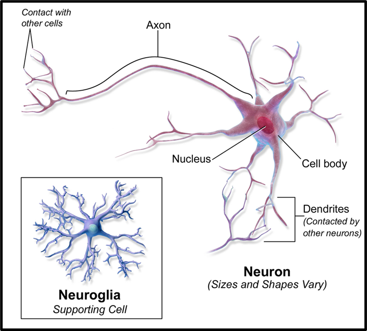
🧩 Components of Neural Tissue
Neural tissue consists of two major cell types:
1️⃣ Neurons (Functional Units)
Neurons are excitable cells that transmit signals.
Structure of a Neuron:
| Part | Function |
| Cell Body | Contains nucleus & Nissl’s granules |
| Dendrites | Receive signals |
| Axon | Transmits signals away |
👉 Signal direction:
Dendrite → Cell body → Axon
Myelin Sheath
- A lipid-protein covering around axons
- Formed by:
- Schwann cells (Peripheral Nervous System)
- Oligodendrocytes (Central Nervous System)
👉 Function:
- Acts as insulation
- Increases speed of impulse transmission
2️⃣ Neuroglia (Supporting Cells)
These are not involved in signal transmission directly but are essential for neuron survival.
| Type | Function |
| Astrocytes | Structural support, blood-brain barrier |
| Microglia | Defense (phagocytosis) |
| Oligodendrocytes | Myelin in CNS |
| Schwann cells | Myelin in PNS |
| Ependymal cells | Produce cerebrospinal fluid (CSF) |
👉 Neuroglia form more than half of neural tissue volume
🔄 Synapse (Key Concept)
A synapse is the junction where → Axon of one neuron meets dendrite of another neuron
👉 This is where information transfer occurs (chemical + electrical signals)
⚖️ Axon vs Dendrite
| Feature | Axon | Dendrite |
| Structure | Long, single | Short, branched |
| Function | Sends signals | Receives signals |
| Number | Usually one | Multiple |
| Myelin | Often present | Rare |
🧠 Functions of Neural Tissue
Neural tissue acts like the command and control system of the body:
- Signal transmission → connects body parts
- Information processing → decision-making
- Motor control → voluntary & involuntary actions
- Homeostasis → regulates internal balance
- Cognition → thinking, memory, learning
- Sensory input → receives environmental signals
Vascular and Avascular Tissue in Animals
Basic Definition
- Vascular Tissues: Tissues that contain blood vessels (arteries, veins, capillaries) for direct supply of nutrients and oxygen.
- Avascular Tissues: Tissues that lack blood vessels; nourishment occurs indirectly through diffusion.
Comparison
| Feature | Vascular Tissue | Avascular Tissue |
| Definition | Contains blood vessels for supply of nutrients and oxygen | Lacks blood vessels |
| Nutrient & Oxygen Supply | Direct supply via capillaries, arteries, and veins | Indirect supply through diffusion from nearby tissues |
| Waste Removal | Efficient via bloodstream | Slower, depends on surrounding tissues |
| Healing Ability | Rapid healing due to rich blood supply | Slow healing due to absence of direct blood flow |
| Structure | Complex with organised vascular network | Simple, lacks vascular components |
| Function | Supports high metabolic activity, growth, and repair | Provides structural roles and reduces friction (e.g., joints) |
| Flexibility | Generally, less flexible (e.g., bone) | More flexible (e.g., cartilage) |
| Examples | Bones, muscles, skin, internal organs | Cartilage, cornea, lens, epidermis |