
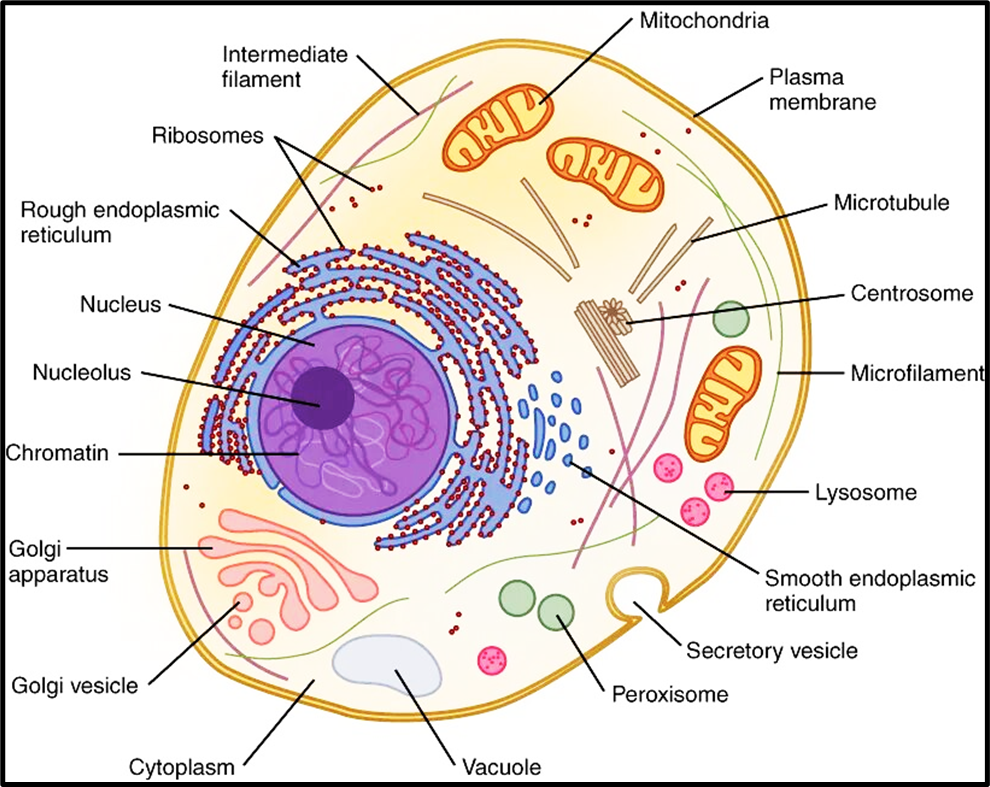
Cell and its Organelles
Think of a cell as a bustling city. Every city has specialised departments — the power plant, the waste disposal unit, the communication tower. Similarly, a cell has cell organelles — each a specialised structure performing a distinct function necessary for the cell’s survival.
🚪 The Cell Membrane (Plasma Membrane) — The Intelligent Gatekeeper
The cell membrane is the outermost covering of the cell that separates its internal contents from the external environment. But calling it merely a “boundary wall” would be a gross understatement. It is dynamic, intelligent, and multifunctional.
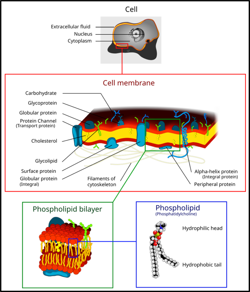
Composition — What is it made of?
The membrane is built like a sandwich — and understanding this sandwich is critical. Three components make up the cell membrane:
1. Lipid bilayer — Two layers of phospholipids, each having a hydrophilic (water-loving) head and a hydrophobic (water-fearing) tail (see diagram). The tails point inward (away from water), heads point outward. This clever arrangement is what makes the membrane selectively permeable — it doesn’t let everything through.
2. Proteins — Two types:
- Integral proteins — embedded deep, spanning the entire membrane. They act as channels and carriers.
- Peripheral proteins — sitting on the surface, involved in signalling and structural support.
3. Carbohydrates — Attached to proteins (glycoproteins) or lipids (glycolipids) on the outer surface. They serve as identification tags — like name badges on a cell — crucial for immune recognition.
The Fluid Mosaic Model (Singer & Nicolson, 1972) explains this structure. “Fluid” because the lipid layer is quasi-fluid and proteins can move laterally. “Mosaic” because proteins are scattered across the membrane like tiles in a mosaic.
Functions of the Cell Membrane
The cell membrane is not passive — it is actively managing the cell’s interaction with the world:
- Selective permeability — lets nutrients and oxygen in, keeps harmful substances out, removes waste.
- Protection and structural support — works with the cytoskeleton to maintain cell shape and integrity.
- Cellular communication — receptor proteins detect signals like hormones, influencing growth, metabolism, and immune responses.
- Transport of molecules — via transport proteins, moving ions and nutrients by both passive and active mechanisms.
- Cell recognition — glycoproteins and glycolipids enable cells to identify each other. Critical for immune responses, tissue formation, and adhesion.
- Homeostasis — maintains a stable internal environment by controlling what enters and exits.
- Endocytosis, exocytosis, cell division — the membrane’s fluidity enables all these dynamic cellular functions.
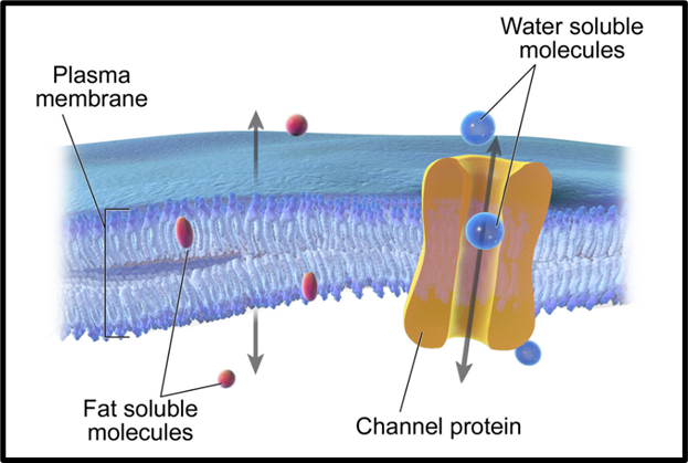
🚗 Transport Mechanisms — How Things Move Across the Membrane
1. Diffusion — Nature’s Equaliser
Diffusion is the movement of molecules from an area of higher concentration to an area of lower concentration — until they are evenly distributed. No energy is required, so it is passive transport.
Classic example: When you open a perfume bottle, the molecules spread across the room — from high concentration (bottle) to low concentration (room). In biology, this is how O₂ and CO₂ are exchanged in the alveoli of the lungs.
UPSC note: In pneumonia, alveoli fill with pus and fluid, disrupting this diffusion — that’s why breathing becomes difficult.
Selective permeability of the plasma membrane allows fat-soluble molecules to diffuse directly while water-soluble molecules require channel proteins for transport.
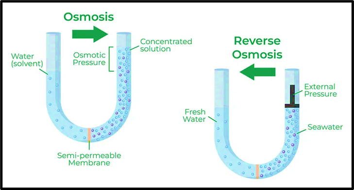
2. Osmosis — Water’s Version of Diffusion
Osmosis is essentially diffusion, but exclusively for water (the solvent), across a semi-permeable membrane.
The rule is simple: water moves from where there are fewer dissolved particles (low solute concentration) to where there are more dissolved particles (high solute concentration) — to equalise concentrations on both sides.
Think of it this way — if you put a raisin in water, it swells up. The raisin’s interior has higher solute concentration than plain water, so water rushes in via osmosis. Conversely, if you soak it in concentrated salt solution, it shrivels.
Solute = the substance dissolved (e.g., salt). Solvent = the substance doing the dissolving (e.g., water).
Plant roots absorb water from soil through osmosis. Unicellular freshwater organisms also gain water this way.
Reverse Osmosis (RO) — Technology Fights Nature
In normal osmosis, water flows naturally from pure side to impure side. Reverse Osmosis does the opposite — by applying external pressure greater than the osmotic pressure, it forces water from the impure side to the pure side, leaving contaminants behind.
Osmotic pressure = the minimum pressure needed to stop natural osmosis from occurring.
The comparative table below captures the key distinctions:
| Feature | Osmosis | Reverse Osmosis |
| Direction | Pure → Impure | Impure → Pure |
| Driving force | Osmotic pressure (natural) | Applied pressure (artificial) |
| Process type | Natural | Technology |
| Example | Plant roots absorbing water | Drinking water purification, desalination |
Advantages of RO: High purification, removes bacteria and viruses, softens hard water by removing Ca²⁺ and Mg²⁺ (scale-causing ions).
Disadvantages of RO: Removes beneficial minerals (Na, Ca, Mg, K), high energy consumption, water wastage, slow process, costly installation, and complex maintenance.
Osmosis is the natural movement of water from dilute to concentrated solution across a semi-permeable membrane, whereas reverse osmosis uses external pressure to force water in the opposite direction for purification.
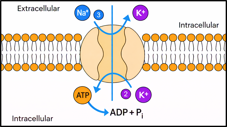
3. Active Transport — Going Against the Current
Sometimes the cell needs to move substances against the concentration gradient — from low to high concentration. This is like pushing water uphill — it requires energy in the form of ATP.
The classic example is the Na⁺/K⁺ pump — a protein that pumps sodium ions out of the cell and potassium ions in, even though the natural concentration gradient would push them the other way. This is critical for nerve impulse transmission.
4. Endocytosis and Exocytosis — Bulk Transport
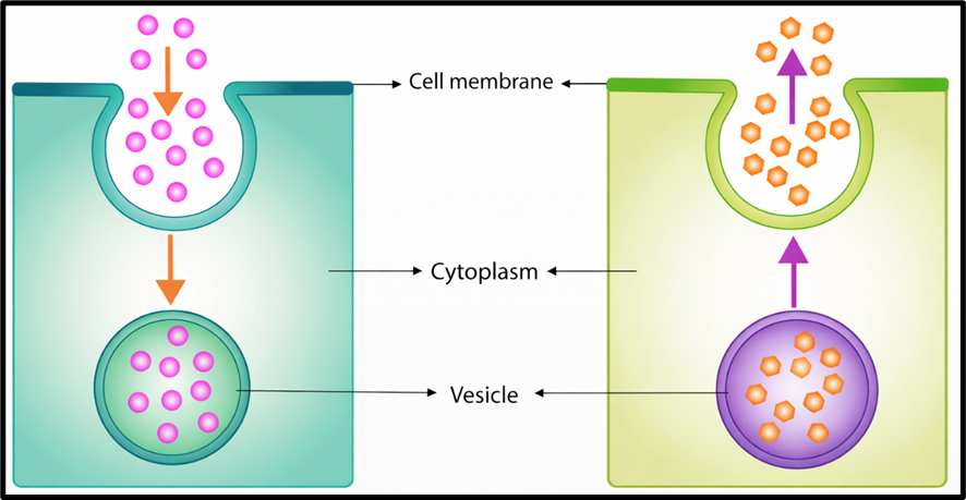
Sometimes substances are too large to pass through transport proteins. The cell then uses vesicle-based mechanisms:
- Endocytosis — the cell membrane folds inward to engulf external material, forming a vesicle that enters the cell. Think of it as the cell “eating” or “drinking.” Amoeba captures its food this way.
- Exocytosis — a vesicle inside the cell fuses with the cell membrane and releases its contents outside. Think of it as the cell “spitting out.” Hormones and neurotransmitters are secreted this way.
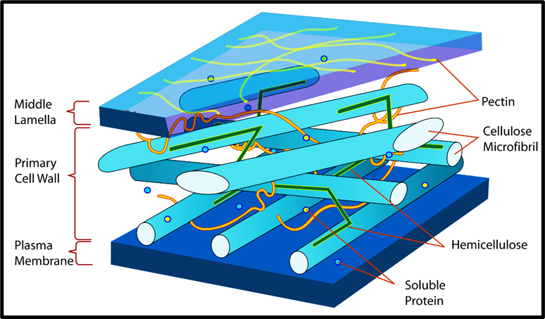
🧱 Cell Wall
If the cell membrane is the intelligent gatekeeper, the cell wall is the fort wall built around it — rigid, strong, and non-living. Crucially, it is found only in plants, bacteria, fungi, algae, and archaea — not in animal cells.
Think of it this way to remember: animals can move away from danger, so they don’t need rigid walls. Plants, bacteria, and fungi are stationary — they need structural armour.
Composition — The Fort is Built Differently for Different Kingdoms!
Each organism type has a unique cell wall composition:
| Organism | Cell Wall Composition |
| Plants | Cellulose, hemicellulose, pectins, proteins |
| Algae | Cellulose, galactans, mannans, calcium carbonate |
| Fungi | Glucans, chitin, glycoproteins |
| Bacteria | Peptidoglycan (murein) |
| Archaea | Polysaccharides and glycoconjugates |
UPSC trick:
Fungi = Chitin (same material as insect exoskeletons — surprisingly!).
Bacteria = Peptidoglycan — this is why antibiotics like penicillin work by targeting peptidoglycan synthesis.
No peptidoglycan = bacteria burst and die.
Functions of the Cell Wall
- Structural support and shape — gives rigidity to stems, leaves; holds the cell’s form against physical forces.
- Protection — shields against mechanical stress (temperature, wind, moisture), osmotic pressure, and pathogens (bacteria, fungi, viruses).
- Regulation of cell growth — controls the direction and rate at which the cell grows.
- Selective permeability — allows specific molecules and ions to pass, helping regulate the internal environment.
- Communication via plasmodesmata — channels that pass through the cell wall, connecting the cytoplasm of neighbouring cells. Think of them as underground tunnels between adjacent buildings, allowing exchange of materials and signalling molecules.
Middle lamella — a layer mainly of calcium pectate that acts like glue between neighbouring cells, holding them together. This is why overcooking vegetables makes them mushy — heat dissolves the calcium pectate!
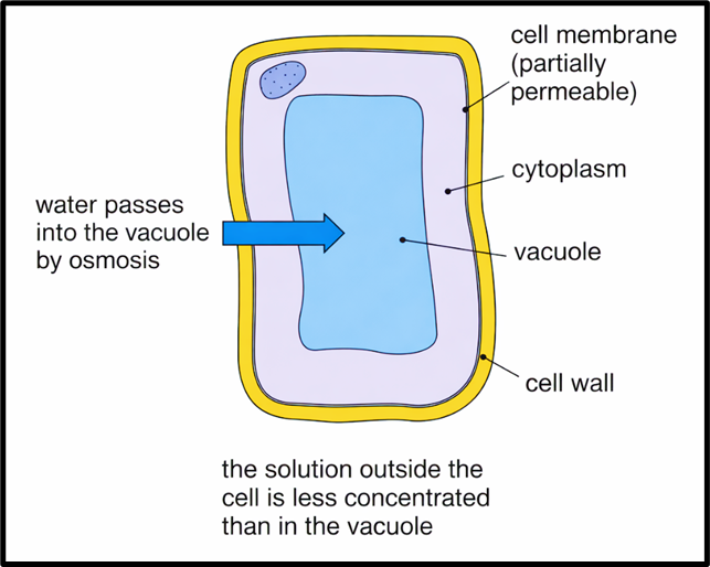
🌊 Turgor Pressure — The Secret Behind Why Plants Stand Tall
This is one of the most elegant concepts in plant biology.
Imagine you fill a balloon with water and press it against a rigid wall. The balloon pushes outward, the wall pushes back — and the whole system becomes taut and firm. This is exactly what happens in a plant cell.
When a plant cell is in a hypotonic environment (solute concentration outside < solute concentration inside), water enters the cell by osmosis. The cell swells and pushes against the rigid cell wall. The cell wall pushes back with equal force. This internal pressure of fluid pressing the cell membrane against the cell wall is called turgor pressure (also called hydrostatic pressure).
Benefits of turgor pressure — three critical functions:
- Maintaining structure — keeps plant cells firm and upright. Loss of turgor = wilting.
- Plant growth — turgor pressure causes cell elongation by pushing the cell wall outward as water enters, making the cell grow longer.
- Stomatal movement — the opening and closing of stomata (tiny pores on plant surfaces for gas exchange) is directly controlled by turgor pressure changes in guard cells.
🧠 Nucleus — The Brain of the Cell
Robert Brown discovered the nucleus. It is a membrane-bound organelle found in eukaryotic cells — generally spherical, located at the centre, and housing the cell’s genetic material, DNA. It is rightly called the “brain of the cell” — it directs reproduction, development, and all metabolic activities.
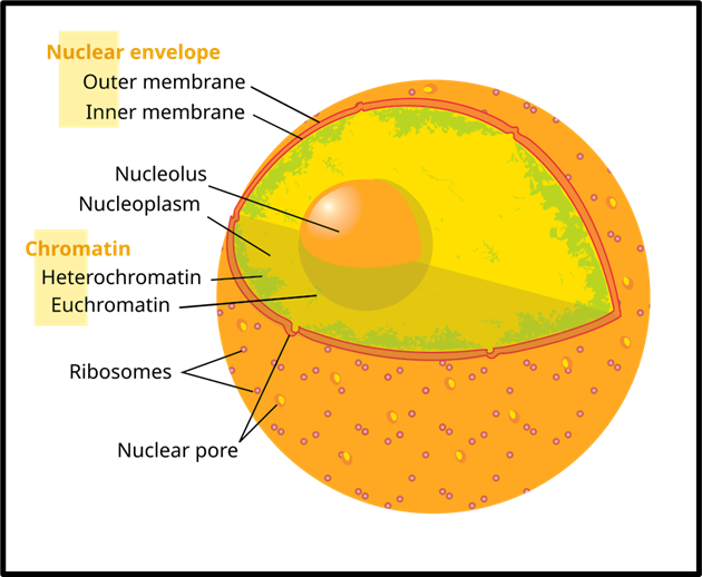
Composition of the Nucleus
Nuclear Membrane (Nuclear Envelope) A double-membrane structure enclosing the nucleus and separating it from the cytoplasm. It has nuclear pores — regulated gateways that control the exchange of materials (like RNA going out, proteins coming in) between the nucleus and cytoplasm.
Nucleoplasm (Nuclear Matrix) The fluid-like interior of the nucleus, containing two critical structures:
- Nucleolus — a dense, spherical body within the nucleoplasm. Its primary job is ribosomal RNA (rRNA) synthesis — manufacturing the raw material needed to build ribosomes, which in turn make proteins. Think of it as the factory that builds the protein-making machines.
- Chromosomes — thread-like structures made of DNA coiled around histone proteins. Each chromosome carries genes — the functional segments of DNA that carry instructions for an organism’s development, function, and inheritance. Humans have 46 chromosomes (23 pairs).
🌊 Cytoplasm
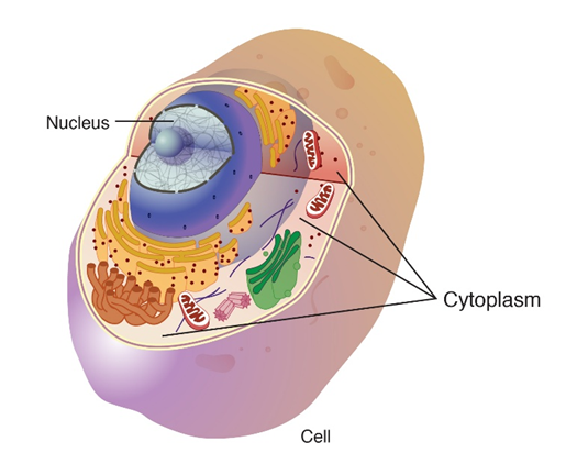
The cytoplasm is the jelly-like substance filling the space between the nucleus and the cell membrane. It consists of:
- Cytosol — the fluid component (mostly water with dissolved salts and molecules)
- Cell organelles — mitochondria, endoplasmic reticulum, Golgi apparatus, and others (we’ll explore these in the next section!)
- Various particles — ribosomes, vesicles, inclusions
The cytoplasm is not merely a passive filler. It maintains cell shape, facilitates movement of materials within the cell, and is the site of many biochemical reactions.
Protoplasm = Cytoplasm + Nucleus. The term was coined by Purkinje in 1839. It refers to the entire living content of the cell. Think of it as the “total living matter” of a cell.
🏭 The Endomembrane System
The endomembrane system is a network of interconnected organelles and membranes within a eukaryotic cell whose functions are coordinated with each other. It includes: the nuclear envelope, endoplasmic reticulum (ER), Golgi apparatus, lysosomes, vacuoles, vesicles, and plasma membrane.
Important UPSC distinction: Mitochondria, chloroplasts, and peroxisomes are NOT part of the endomembrane system — because their functions are independent and not coordinated with the system’s components.
🏗️ Endoplasmic Reticulum (ER) — The Cell’s Highway + Factory
The ER is an extensive network of membrane-bound tubes and sheets — imagine a vast internal highway system running through the cell. There are two types:
- Rough ER — has ribosomes attached to its surface, giving it a rough appearance. Ribosomes are the actual sites of protein synthesis. The rough ER is essentially the manufacturing floor of the cell.
- Smooth ER — lacks ribosomes. It is responsible for lipid (fat) synthesis and, very importantly, detoxification — especially in liver cells of vertebrates, where it breaks down drugs and poisons.
Some proteins and lipids produced here contribute to membrane biogenesis — the building of new cell membranes. Others function as enzymes and hormones.
📦 Golgi Apparatus
Described first by Camillo Golgi, this organelle consists of stacked, flattened membrane sacs called cisterns. Think of it as the post office and quality control centre of the cell.
Proteins and lipids produced in the ER arrive here in vesicles, get modified, packaged, and sorted, then are dispatched to their correct destinations — whether to the cell membrane, outside the cell, or to lysosomes.
Three key functions:
- Delivery of proteins and lipids — transports, modifies, and packages them into vesicles for targeted delivery.
- Complex sugar synthesis — converts simple sugars into complex ones.
- Lysosome formation — creates lysosomes, the cell’s digestion and waste management units.
💣 Lysosomes — The Suicide Bags of the Cell
Lysosomes are membrane-bound sacs packed with powerful digestive enzymes produced by the rough ER. They are the cell’s sanitation and demolition squad.
Functions:
- Digestion — break down macromolecules, old cell parts, and foreign invaders (bacteria, food particles) into simpler molecules.
- Waste removal — enzymatically eliminate cellular debris.
- Autophagy — recycle damaged organelles. The cell essentially eats itself to reclaim useful components.
- Apoptosis (programmed cell death) — when a cell needs to die (e.g., during embryo development, or when infected), lysosomes rupture and release their digestive enzymes into the cytoplasm, destroying the cell from within. This is why they are famously called “suicide bags” of the cell.
🪣 Vacuoles — The Cell’s Storage Tanks
Vacuoles are membrane-bound storage compartments in the cytoplasm. They store water, sap, amino acids, sugars, organic acids, proteins, and excretory products. They are bounded by a single membrane called the tonoplast.
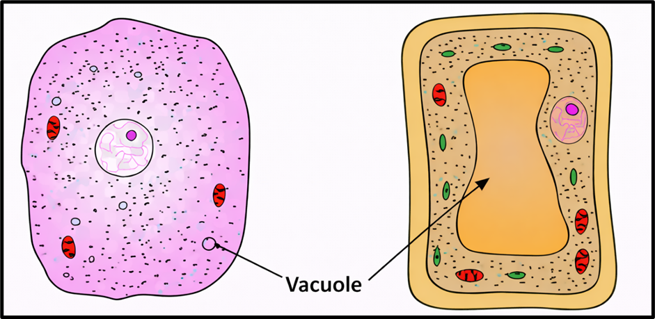
Key comparisons:
- Animal cells — vacuoles are small and temporary.
- Plant cells — vacuoles are enormous, potentially occupying 50–90% of the cell volume. This central vacuole filled with cell sap is what gives plant cells their turgidity — directly linked to turgor pressure discussed earlier!
Special types:
- Contractile vacuole in Amoeba — vital for osmoregulation (regulating water balance) and excretion.
- Food vacuoles in protists — formed by engulfing food particles via endocytosis.
⚡ Mitochondria — The Powerhouse of the Cell
This is perhaps the most famous organelle name in biology — and for good reason!
Mitochondria are sausage or cylindrically shaped, double membrane-bound structures. The outer membrane forms a smooth boundary. The inner membrane folds inward into structures called cristae — these folds dramatically increase the surface area available for chemical reactions. The inner compartment is filled with a dense fluid called the matrix.
The mitochondria convert glucose + oxygen → ATP through cellular respiration. ATP (adenosine triphosphate) is the energy currency of the cell — every cellular process that needs energy draws from this account.
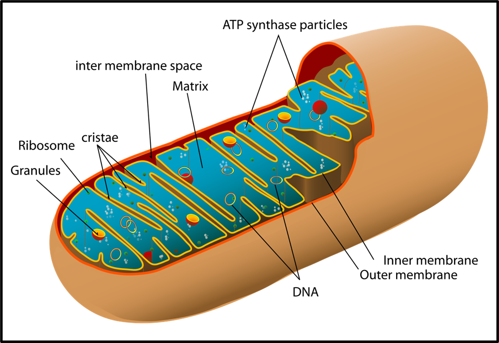
Two remarkable facts about mitochondria:
- They have their own DNA and ribosomes — meaning they can synthesise some of their own proteins independently. This is evidence for the endosymbiotic theory — the idea that mitochondria were once free-living bacteria that got incorporated into larger cells billions of years ago.
- They divide by fission — like bacteria do — further supporting their bacterial ancestry.
🌿 Plastids — Exclusive to Plants and Euglenoids
Plastids are found only in plant cells and euglenoids. They contain specific pigments and are classified into three types based on those pigments:
| Plastid | Pigment | Colour | Function |
| Chloroplasts | Chlorophyll + carotenoids | Green | Photosynthesis |
| Chromoplasts | Carotene, xanthophylls | Yellow/Orange/Red | Attract pollinators |
| Leucoplasts | None (colourless) | — | Storage (starch/oil/protein) |
Leucoplast subtypes — Amyloplasts store starch (potatoes!), elaioplasts store oils/fats, aleuroplasts store proteins.
Inside the Chloroplast
The chloroplast is double-membraned. The space inside the inner membrane is called the stroma — containing enzymes for carbohydrate and protein synthesis, DNA, and ribosomes.
Within the stroma sit flattened membrane sacs called thylakoids, stacked in columns called grana (singular: granum), connected by stroma lamellae. The chlorophyll pigments sit inside the thylakoids — this is where light energy is captured during photosynthesis.
Like mitochondria, chloroplasts also have their own DNA and ribosomes — again supporting the endosymbiotic theory.
⚙️ Ribosomes — The Protein Factories
Ribosomes are small granular structures made of RNA and proteins, first observed by George Palade in 1953. They are the actual sites of protein synthesis — the most fundamental molecular activity in any living cell.
- They have no membrane (unlike most organelles).
- Each ribosome has two subunits: a larger and a smaller one.
- Eukaryotic ribosomes (80S) are larger than prokaryotic ribosomes (70S). The ‘S’ stands for Svedberg unit — a measure of sedimentation rate, indirectly indicating size and density.
UPSC relevance: Many antibiotics (like streptomycin, erythromycin) work by targeting the 70S prokaryotic ribosome — disrupting bacterial protein synthesis without harming human cells (which have 80S ribosomes). This size difference is what makes these antibiotics selectively toxic to bacteria.
🕸️ Cytoskeleton, Cilia, Flagella
Cytoskeleton — a network of protein fibres in the cytoplasm that provides structural support, maintains cell shape, and facilitates movement of materials within the cell. Think of it as the cell’s internal scaffolding and transport tracks.
Cilia and Flagella — hair-like extensions of the cell membrane, both covered by the plasma membrane and having a central core called the axoneme.
| Feature | Cilia | Flagella |
| Length | Short | Long |
| Number | Numerous | Few |
| Movement | Oar-like, moves surrounding fluid | Propels the whole cell |
| Example | Respiratory tract, fallopian tubes | Sperm cells, bacteria |
Microvilli vs Cilia — a commonly confused pair:
| Feature | Microvilli | Cilia |
| Structure | Finger-like projections | Hair-like projections |
| Function | Increase surface area for absorption | Move fluids/particles across surface |
| Motility | Non-motile | Motile |
| Location | Small intestine, kidney tubules | Respiratory tract, fallopian tubes |
🔵 Centrosome and Centrioles — The Cell Division Organisers
A centrosome contains two cylindrical structures called centrioles. Their two roles are:
- Form the basal body of cilia and flagella.
- During cell division in animal cells, they generate spindle fibres that form the spindle apparatus — the machinery that pulls chromosomes apart during division.
Notably, plant cells generally lack centrioles — they form spindle fibres through other mechanisms.