
Excretion in Human Beings
Every living organism continuously performs metabolic activities—breaking down food, synthesizing molecules, and generating energy. During these processes, several waste products are formed such as:
- Ammonia, urea, uric acid (nitrogenous wastes)
- Carbon dioxide
- Excess water and ions (Na⁺, K⁺, Cl⁻)
Now, if these wastes accumulate in the body, they become toxic and disturb internal balance. Therefore, the body must eliminate them. This process is called excretion.
Nitrogenous Wastes: The Core Concept
The most important wastes to understand are nitrogenous wastes, which arise from protein metabolism. There are three major types, and you should remember them in terms of toxicity and water requirement:
(1) Ammonia
- Most toxic | Highly water-soluble | Requires large amounts of water for elimination
(2) Urea
- Moderately toxic | Requires less water | Main waste in humans and mammals
(3) Uric Acid
- Least toxic | Requires minimal water | Can be excreted as semi-solid paste
👉 So, there is a clear evolutionary trend:
Ammonia → Urea → Uric Acid (decreasing toxicity and water requirement)
Types of Excretion Based on Nitrogenous Waste
Depending on which waste an organism excretes, we classify them into three categories:
(A) Ammonotelism
- Excretion of ammonia
- Seen in → Aquatic animals (bony fishes, amphibians, aquatic insects)
- Mechanism → Ammonia diffuses directly through gills or body surface
- Logic → Since water is abundant, toxicity is diluted easily
(B) Ureotelism
- Excretion of urea
- Seen in:
- Mammals (including humans)
- Many terrestrial amphibians
- Some marine fishes
- Process:
- Ammonia is converted into urea in the liver
- Urea is filtered by kidneys and excreted in urine
- Special point:
- Some urea is retained to maintain osmolarity (water balance)
(C) Uricotelism
- Excretion of uric acid
- Seen in → Reptiles, birds, insects, land snails
- Advantage:
- Excreted as solid or paste
- Helps in water conservation
- Evolutionary significance → Very useful for terrestrial life, especially in dry environments
Excretory Structures in Different Organisms
Now, nature has designed different structures for excretion depending on the complexity of organisms.
A. Invertebrates (Simple Animals)
These organisms have relatively simple excretory systems:
- Flame Cells (Protonephridia)
- Found in flatworms
- Function → Excretion + osmoregulation (water balance)
- Nephridia
- Found in earthworms (annelids)
- Function:
- Remove nitrogenous waste
- Maintain internal fluid balance
- Malpighian Tubules
- Found in insects (e.g., cockroach)
- Function:
- Remove waste directly from body fluids
- Help in water conservation
- Green Glands (Antennal Glands)
- Found in crustaceans (e.g., prawns)
- Function → Excretion and ionic balance
B. Vertebrates (Complex Animals)
In higher organisms like humans, excretion is carried out by highly specialized organs:
Kidneys
- Main excretory organs
- Functions:
- Filter blood
- Remove urea and other wastes
- Maintain water balance
- Regulate electrolytes (Na⁺, K⁺, Cl⁻)
This regulation of water and salts is called osmoregulation, which is crucial for maintaining internal stability (homeostasis).
Components of the Human Excretory System
The system is composed of four main parts, working in a coordinated manner:
- Kidneys → Filter blood and form urine
- Ureters → Transport urine from kidneys to bladder
- Urinary bladder → Temporary storage of urine
- Urethra → Passage for elimination of urine
Think of it like a filtration–transport–storage–discharge system.
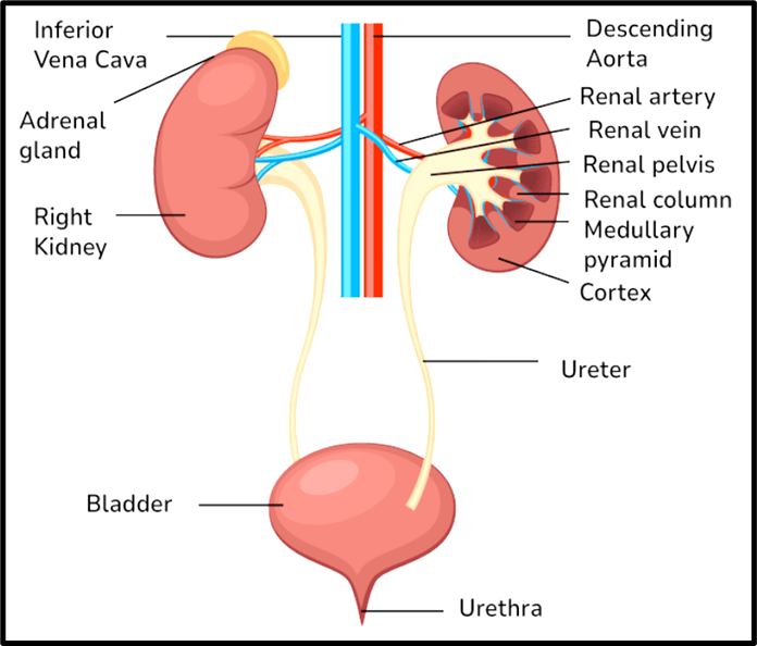
Structure of the Kidneys
The kidneys are paired, reddish-brown, bean-shaped organs located towards the back of the abdomen. Each kidney is about 10–12 cm long, which gives you an idea of how compact yet efficient they are.
Key Structural Features
- On the inner side, there is a depression called the hilum, where:
- Blood vessels enter and leave
- Ureter emerges
- Nerves pass
- Inside the hilum lies the renal pelvis, a funnel-shaped cavity that collects urine. It further branches into calyces.
- The kidney is protected by a tough capsule.
Internal Organization
The kidney is divided into two major zones:
(A) Cortex (Outer Region)
- Extends inward as renal columns (Columns of Bertini)
- Contains major portions of nephrons
(B) Medulla (Inner Region)
- Organized into cone-shaped medullary pyramids
- These project into calyces and play a key role in urine concentration
Nephron: The Functional Unit
Each kidney contains approximately 1 million nephrons, making it a highly efficient filtration system.
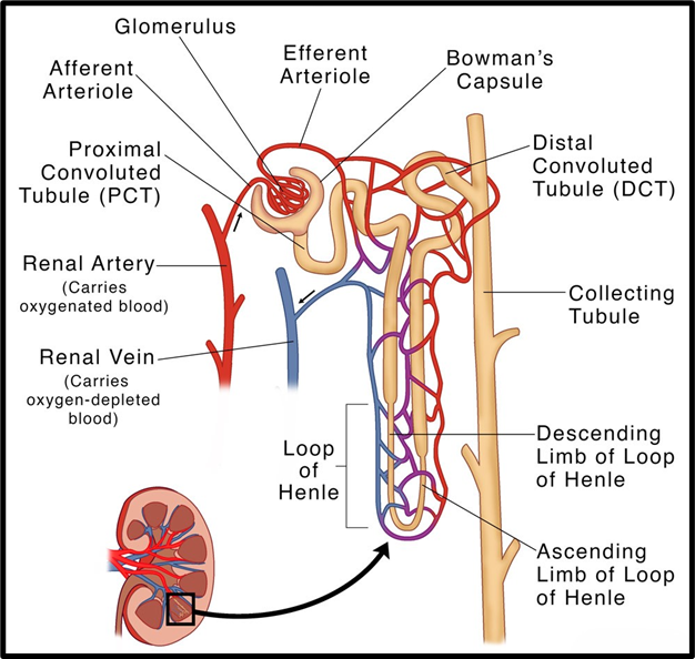
A nephron has two main parts:
(A) Glomerulus
- A network of capillaries formed by the afferent arteriole
- Blood exits through the efferent arteriole
- Primary function: filtration of blood
(B) Renal Tubule
This is where the real “processing” happens. It has multiple segments:
- Bowman’s Capsule
- Cup-shaped structure enclosing the glomerulus
- Together they form the Malpighian body (renal corpuscle)
- Proximal Convoluted Tubule (PCT)
- Loop of Henle
- Descending limb
- Ascending limb
- Distal Convoluted Tubule (DCT)
- Collecting Duct → Final urine collection
Types of Nephrons
- Cortical nephrons
- Short Henle’s loop
- Mainly in cortex
- Juxtamedullary nephrons
- Long loops extending deep into medulla
- Crucial for urine concentration
Special Capillary System
- After filtration, the efferent arteriole forms:
- Peritubular capillaries → Surround tubules
- Vasa recta → U-shaped vessels (important for concentration mechanism)
Urine Formation
Urine formation is not a single step—it is a three-stage process, happening sequentially in different parts of the nephron:
(1) Glomerular Filtration (Ultrafiltration)
- Blood is filtered in the glomerulus due to high pressure
- Filtration rate:
- ~ 125 ml/min
- ~ 180 litres/day
- Almost everything except proteins passes into the filtrate
- This rate is called Glomerular Filtration Rate (GFR)
- Regulation:
- Controlled by Juxtaglomerular Apparatus (JGA)
- Releases renin if GFR falls → increases blood flow
👉 Key idea: The body filters a huge volume, but most of it is reclaimed later.
(2) Reabsorption
Out of 180 litres filtered, only about 1.5 litres becomes urine. This means ~99% is reabsorbed.
Types of Reabsorption
- Active transport
- Requires energy
- Reabsorbs: glucose, amino acids, Na⁺
- Passive transport
- No energy required
- Reabsorbs: water, some wastes
(3) Tubular Secretion
- Certain substances are actively added into the filtrate → H⁺ ions, K⁺ ions, Ammonia
👉 Purpose:
- Maintain acid-base balance
- Regulate ionic composition
Functional Role of Different Tubular Segments
Now comes the most important conceptual clarity—each part of nephron has a specific role:
(A) Proximal Convoluted Tubule (PCT)
- Major site of reabsorption (70–80%)
- Reabsorbs → Water, Electrolytes, Glucose, amino acids
- Also secretes → H⁺, NH₃, K⁺
👉 Role: Bulk recovery + pH regulation
(B) Loop of Henle
This is the counter-current system, crucial for concentration:
- Descending limb
- Permeable to water
- Water leaves → filtrate becomes concentrated
- Ascending limb
- Impermeable to water
- Electrolytes move out → filtrate becomes dilute
👉 Role: Creates osmotic gradient in medulla
(C) Distal Convoluted Tubule (DCT)
- Fine-tuning segment
- Reabsorbs → Na⁺ and water (as per body needs)
- Secretes → H⁺, K⁺, NH₃
👉 Role: Hormonal regulation + ionic balance
(D) Collecting Duct
- Final concentration of urine
- Reabsorbs → Large amount of water
- Allows → Some urea recycling → maintains osmolarity
- Also regulates → pH, Ionic balance
👉 Role: Determines final urine concentration
Regulation of Kidney Function
The kidneys continuously adjust urine formation based on the body’s needs. This regulation is achieved through a hormonal feedback mechanism involving → Hypothalamus, Juxta-glomerular apparatus (JGA), Heart
Think of it as a three-level control system managing water balance, blood pressure, and ionic equilibrium.
(A) Role of Hypothalamus and ADH
The hypothalamus contains specialized cells called osmoreceptors, which monitor → Blood volume, Fluid levels, Ion concentration
When the body loses water (e.g., dehydration):
- Osmoreceptors detect increased osmolarity
- Hypothalamus releases Antidiuretic Hormone (ADH)
Action of ADH:
- Increases water reabsorption in kidney tubules
- Reduces urine output (anti-diuresis)
- Also causes vasoconstriction, raising blood pressure
When excess water is present:
- ADH secretion is reduced
- More water is excreted → dilute urine
👉 Key idea: ADH maintains water balance and blood pressure simultaneously.
(B) Role of Juxta-Glomerular Apparatus (JGA)
The JGA acts as a pressure sensor in the kidney.
When blood pressure or flow decreases → JG cells release renin
This initiates the Renin–Angiotensin–Aldosterone System (RAAS):
- Renin converts angiotensinogen → angiotensin I
- Angiotensin I → angiotensin II
Functions of Angiotensin II:
- Causes vasoconstriction → increases blood pressure
- Enhances glomerular filtration
- Stimulates release of aldosterone
Role of Aldosterone:
- Increases Na⁺ and water reabsorption
- Raises blood volume and pressure
👉 Key idea: RAAS is a long-term regulator of blood pressure and filtration rate.
(C) Role of Heart: Atrial Natriuretic Factor (ANF)
When blood volume becomes excessive, the heart responds.
Stimulus → Increased blood flow stretches the atria
Response → Release of Atrial Natriuretic Factor (ANF)
Functions of ANF:
- Causes vasodilation
- Lowers blood pressure
- Promotes excretion of sodium and water
👉 Important balance:
- RAAS → increases blood pressure
- ANF → decreases blood pressure
This creates a homeostatic balance system.
Micturition
Once urine is formed, it must be expelled in a controlled manner.
Mechanism:
- Urine accumulates in the urinary bladder
- As it fills, stretch receptors in the bladder wall are activated
- Signals are sent to the Central Nervous System (CNS)
Response:
- Bladder muscles (detrusor muscle) contract
- Urethral sphincter relaxes
- Urine is expelled
This process is called micturition, controlled by the micturition reflex.
Normal Urine Characteristics
- Volume: 1–1.5 litres/day
- Colour: Light yellow
- pH: Slightly acidic (~6.0)
- Contains → 25–30 g of urea
Clinical Indicators
- Glycosuria → glucose in urine (diabetes mellitus)
- Ketonuria → ketone bodies in urine
👉 Urine analysis is a powerful diagnostic tool.
Role of Other Organs in Excretion
Although kidneys are primary organs, other systems also assist:
(A) Lungs
- Remove ~200 ml/day of CO₂ and water vapour
(B) Liver
- Excretes waste via bile, including → Bilirubin, biliverdin, Cholesterol, Drugs and toxins
These are eliminated through the digestive tract.
(C) Skin
Sweat glands
- Secrete → Water, NaCl, Small amounts of urea and lactic acid
- Primary role: thermoregulation, but also minor excretion
Sebaceous glands
- Secrete sebum
- Eliminate → Sterols, hydrocarbons, waxes
(D) Saliva
- Removes small amounts of nitrogenous waste
👉 Key takeaway: Excretion is a multi-organ function, not limited to kidneys.
Disorders of the Excretory System
Understanding disorders helps in linking structure with function.
(1) Uremia
- Accumulation of urea in blood
- Cause: Kidney failure
Treatment:
- Hemodialysis → Artificial filtration of blood
- Kidney transplantation
(2) Renal Calculi (Kidney Stones)
- Formation of crystals (e.g., oxalates) in kidney
- Causes pain and obstruction
(3) Glomerulonephritis
- Inflammation of glomeruli
- Reduces filtration efficiency