
Transportation in Human Beings
In simple terms, transportation in the human body means the continuous movement of essential substances such as oxygen, nutrients, hormones, and waste products from one part of the body to another.
Think of the body as a highly organized city:
- Cells are like citizens
- Nutrients and oxygen are supplies
- Waste products are garbage
To manage all this efficiently, the body uses a well-developed transport network, known as the circulatory system.
Components of the Circulatory System
The circulatory system has three major components, each performing a distinct but coordinated function:
(i) Circulatory Medium
This is the fluid that carries substances → Blood, Lymph
(ii) Blood Vessels
These are the pathways through which blood flows:
- Arteries → carry blood away from the heart
- Veins → bring blood back to the heart
- Capillaries → enable exchange of substances with tissues
(iii) Heart
The heart acts as a pump, ensuring continuous circulation of blood throughout the body.
Why Do We Need Such a System?
All living cells require:
- Oxygen for respiration
- Nutrients for energy and growth
- Removal of waste like carbon dioxide and nitrogenous waste
In simpler organisms like sponges, water itself performs this function. But in complex organisms like humans, blood acts as the specialized transport medium, supported by lymph.
Blood: The Main Circulatory Medium
Blood is a special connective tissue composed of:
- A fluid part → Plasma
- Solid components → Formed elements
Its functions include transporting → Oxygen, Carbon dioxide, Nutrients, Hormones, Waste products
Plasma: The Fluid Matrix
Plasma makes up about 55% of blood and acts as the transport medium within the transport medium.
Composition of Plasma
- 90–92% water → acts as solvent
- 6–8% proteins, mainly:
- Fibrinogen → helps in blood clotting
- Globulins → involved in immunity
- Albumins → maintain osmotic balance
Other Components
- Electrolytes: Na⁺, Ca²⁺, Mg²⁺, Cl⁻
- Nutrients: glucose, amino acids, fats
- Waste products
👉 Important distinction:
- Plasma without clotting factors = Serum
Formed Elements (Cellular Components)
These constitute about 45% of blood and include three types:
(A) Erythrocytes (Red Blood Cells – RBCs)
These are the most abundant cells in blood.
Key Features
- Count: ~5–5.5 million per mm³ (in adult males)
- Shape: Biconcave disc (increases surface area for gas exchange)
- Lack nucleus (in mammals) → more space for haemoglobin
- Contain haemoglobin, an iron-containing protein
Functions
- Transport oxygen from lungs to tissues
- Carry a portion of carbon dioxide back
Life Cycle
- Lifespan: ~120 days
- Destroyed in the spleen (hence called graveyard of RBCs)
(B) Leukocytes (White Blood Cells – WBCs)
These are the defence cells of the body.
Key Features
- Count: 6,000–8,000 per mm³
- Nucleated | Colourless (no haemoglobin) | Short-lived
Types of WBCs
1. Granulocytes (with granules)
- Neutrophils
→ Most abundant; first line of defence
→ Perform phagocytosis (engulf pathogens) - Eosinophils
→ Deal with parasitic infections and allergies - Basophils
→ Rarest type
→ Release:- Histamine & serotonin → cause inflammation
- Heparin → prevents clotting
2. Agranulocytes (without granules)
- Lymphocytes
These are central to immunity:- T cells → cell-mediated immunity (destroy infected/cancer cells)
- B cells → humoral immunity (produce antibodies)
- Natural Killer (NK) cells → kill infected or tumour cells directly, without prior exposure
- Monocytes
→ Largest WBCs
→ Differentiate into macrophages
→ Perform phagocytosis (clean up debris and pathogens)
(C) Platelets (Thrombocytes)
These are cell fragments, not complete cells.
Key Features
- Count: 150,000–350,000 per mm³
- Derived from megakaryocytes in bone marrow
Function
- Essential for blood clotting (coagulation)
- Prevent excessive blood loss during injury
👉 Low platelet count can lead to bleeding disorders
Blood Groups: The Identity of Blood
Although all human blood looks similar, it is chemically different. These differences are based on specific molecules present on the surface of RBCs.
Two major systems are used worldwide:
- ABO Blood Group System
- Rh Blood Group System
ABO Blood Grouping
The ABO system is based on the presence or absence of two antigens on RBCs:
- Antigen A
- Antigen B
👉 Antigens are substances that trigger an immune response.
👉 Antibodies are proteins produced by the body to attack foreign antigens.
Basic Principle
Each blood group has:
- Specific antigens on RBCs
- Opposite antibodies in plasma
| Blood Group | Antigens on RBCs | Antibodies in Plasma | Can Receive From |
| A | A | Anti-B | A, O |
| B | B | Anti-A | B, O |
| AB | A and B | None | A, B, AB, O |
| O | None | Anti-A and Anti-B | O |
Key Concepts
1. Why Matching is Important
If incompatible blood is transfused:
- Antibodies attack foreign antigens
- This leads to agglutination (clumping of RBCs)
- It can be fatal
2. Universal Donor and Recipient
- Universal Donor → Blood Group O
- No A or B antigens → cannot be attacked by recipient antibodies
- Universal Recipient → Blood Group AB
- No anti-A or anti-B antibodies → accepts all blood types
| Donor ➡️ / Recipient ⬇️ | O− | O+ | B− | B+ | A− | A+ | AB− | AB+ |
| O− | ✔ | ✖ | ✖ | ✖ | ✖ | ✖ | ✖ | ✖ |
| O+ | ✔ | ✔ | ✖ | ✖ | ✖ | ✖ | ✖ | ✖ |
| B− | ✔ | ✖ | ✔ | ✖ | ✖ | ✖ | ✖ | ✖ |
| B+ | ✔ | ✔ | ✔ | ✔ | ✖ | ✖ | ✖ | ✖ |
| A− | ✔ | ✖ | ✖ | ✖ | ✔ | ✖ | ✖ | ✖ |
| A+ | ✔ | ✔ | ✖ | ✖ | ✔ | ✔ | ✖ | ✖ |
| AB− | ✔ | ✖ | ✔ | ✖ | ✔ | ✖ | ✔ | ✖ |
| AB+ | ✔ | ✔ | ✔ | ✔ | ✔ | ✔ | ✔ | ✔ |
Rh Blood Grouping
Apart from A and B antigens, another important factor is the Rh antigen (Rh factor).
Classification
- Rh-positive (Rh⁺) → Rh antigen present (~80% people)
- Rh-negative (Rh⁻) → Rh antigen absent
👉 The name comes from its discovery in the rhesus monkey.
Why Rh Matching Matters
If an Rh⁻ person receives Rh⁺ blood:
- Their body recognizes Rh antigen as foreign
- Produces anti-Rh antibodies
- Future transfusions become dangerous
Rh Incompatibility in Pregnancy
This is where theory directly connects with clinical relevance.
Scenario
- Mother → Rh⁻
- Baby → Rh⁺
First Pregnancy
- Placenta keeps maternal and fetal blood separate
- Usually no major issue
- But during delivery, some fetal blood may enter maternal circulation
- Mother starts producing anti-Rh antibodies
Subsequent Pregnancy
Now the problem begins:
- Maternal antibodies cross placenta
- Attack fetal RBCs
- Cause: Anaemia, Jaundice and in Severe cases → death
This condition is called erythroblastosis fetalis.
Prevention
Doctors inject anti-Rh antibodies (anti-D) after first delivery → Prevents mother from forming her own harmful antibodies
Blood Coagulation (Clotting): Body’s Emergency Response
Whenever there is injury, the body must prevent blood loss immediately.
Step-by-Step Process
- Injury occurs
- Platelets and damaged tissues release clotting factors
- These activate a cascade of reactions
Key Conversion Steps
- Prothrombin (inactive) → converted into → Thrombin (active enzyme)
- Thrombin converts → Fibrinogen (soluble) into → Fibrin (insoluble threads)
👉 These fibrin threads form a mesh that traps blood cells → Forms a clot (scab)
Important Points
- Calcium ions (Ca²⁺) are essential for clotting
- Platelets play a central role
- The clot prevents excessive bleeding and infection
Lymph
Alongside blood, the body has another transport fluid called lymph.
What is Lymph?
Lymph is formed when:
- Plasma leaks out of capillaries into tissue spaces
- This fluid becomes interstitial (tissue) fluid
- It is then collected by the lymphatic system
Composition
- Similar to plasma but:
- Lacks large proteins
- No RBCs
- Contains lymphocytes (important for immunity)
Functions of Lymph
- Exchange Medium: Transfers nutrients, gases, and waste between blood and tissues
- Immune Defence: Lymphocytes help fight infections
- Fat Transport: Absorbs and transports digested fats from intestines
- Fluid Balance: Returns excess tissue fluid back to blood; Prevents swelling (edema)
Basic Structure of Blood Vessels
Both arteries and veins share a common structural blueprint of three layers, but their thickness and function differ according to the pressure they handle:
- Tunica intima (inner layer)
This is made of smooth endothelium, providing a frictionless surface so blood can flow easily. - Tunica media (middle layer)
Composed of smooth muscle and elastic fibres, this layer is responsible for contraction and relaxation. It is much thicker in arteries because they face high pressure. - Tunica externa (outer layer)
Made of fibrous connective tissue with collagen, it provides structural support and protection.
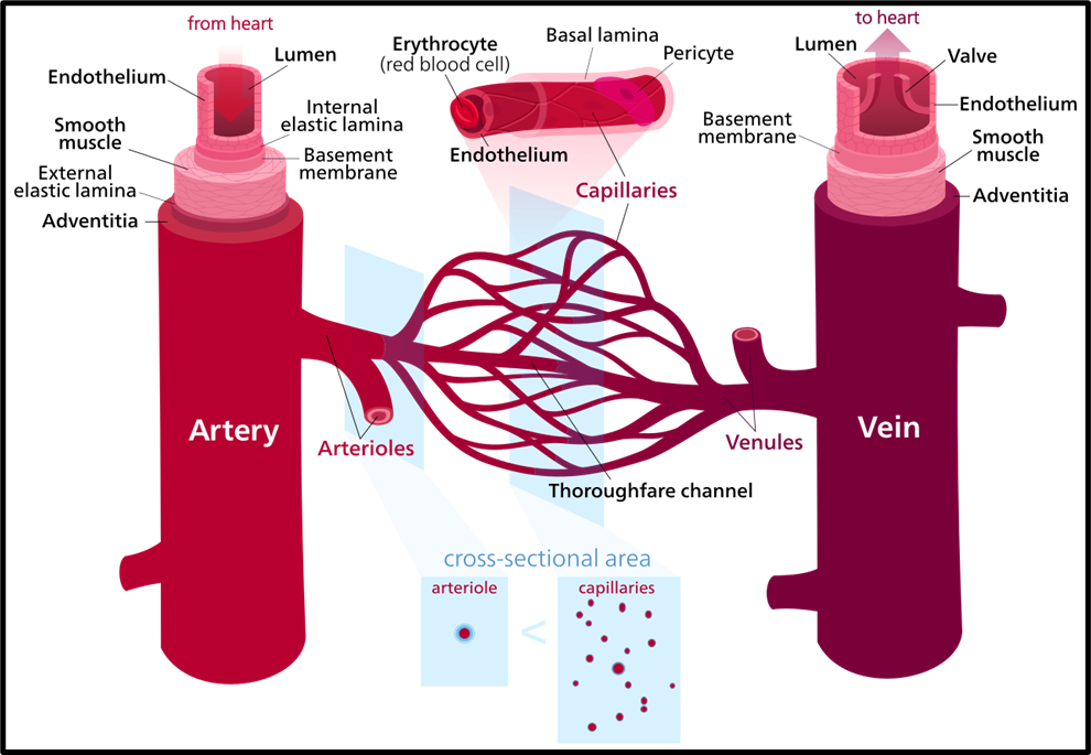
Veins: The Return Pathways
Veins are responsible for bringing blood back to the heart. In most cases, this blood is deoxygenated, except in the pulmonary veins, which carry oxygenated blood from the lungs.
Now, why are veins structurally different?
- Blood returning to the heart is under low pressure, so veins have:
- Thinner walls
- Less muscular and elastic tissue
- To compensate for low pressure, veins contain one-way valves. These valves ensure that blood flows in only one direction—towards the heart—preventing backflow, especially against gravity.
- The smallest veins, called venules, collect blood from capillaries and gradually merge into larger veins.
- Ultimately, major veins like the superior and inferior vena cava carry blood into the right atrium of the heart.
👉 So, veins are not just passive pipes—they actively ensure unidirectional flow under low pressure conditions.
Arteries: The High-Pressure Carriers
Arteries carry blood away from the heart to different parts of the body. This blood is usually oxygenated, except in pulmonary arteries.
Because they directly receive blood pumped by the heart, arteries must withstand high pressure.
Therefore, they have:
- Thick, muscular walls
- High elasticity to expand and recoil with each heartbeat
The largest artery in the body is the aorta, which branches into smaller arteries and then into arterioles, eventually leading to capillaries.
Pulse and Arteries
When the heart pumps blood, arteries expand and recoil. This rhythmic expansion is what we feel as a pulse in places like the wrist or neck.
- The pulse rate reflects the number of heartbeats per minute.
- A normal resting pulse is around 72–80 beats per minute.
👉 Thus, arteries are dynamic vessels, constantly adjusting to maintain efficient blood distribution under high pressure.
Capillaries: The Exchange Interface
Capillaries are the smallest and most delicate blood vessels, but they are the most crucial for survival.
- They connect arterioles (from arteries) to venules (leading to veins).
- Their walls are just one cell thick, which allows:
- Diffusion of oxygen and carbon dioxide
- Exchange of nutrients and waste products
Capillaries form dense networks called capillary beds, ensuring that every cell in the body is in close proximity to blood supply.
👉 In simple terms, arteries and veins transport blood, but capillaries perform the actual exchange that sustains life.
Clotting and Vessel Integrity
If a blood vessel is damaged and starts leaking, platelets quickly act to form a clot.
This prevents:
- Excessive blood loss
- Drop in blood pressure
This mechanism is vital for maintaining the stability of the circulatory system.
Conceptual Understanding
Let’s have an integrated understanding:
- Function:
Arteries distribute blood → Capillaries exchange materials → Veins return blood - Pressure:
Highest in arteries → drops in capillaries → lowest in veins - Wall Thickness:
Thick in arteries (to handle pressure) → extremely thin in capillaries (for exchange) → thin in veins (low pressure) - Valves:
Needed only in veins because blood must move against gravity and low pressure - Pulse:
Felt only in arteries due to direct connection with heart pumping - Location:
Arteries are deeper (protected due to high pressure), veins are often superficial, capillaries are embedded within tissues
If you visualize the system as a continuous loop:
Heart → Arteries → Arterioles → Capillaries → Venules → Veins → Heart
Heart
The heart is a mesodermal organ located in the thoracic cavity, between the lungs, slightly tilted to the left. Its size is roughly that of a clenched fist. It is enclosed in a protective double-walled membrane called the pericardium, which contains pericardial fluid to reduce friction during heartbeats.
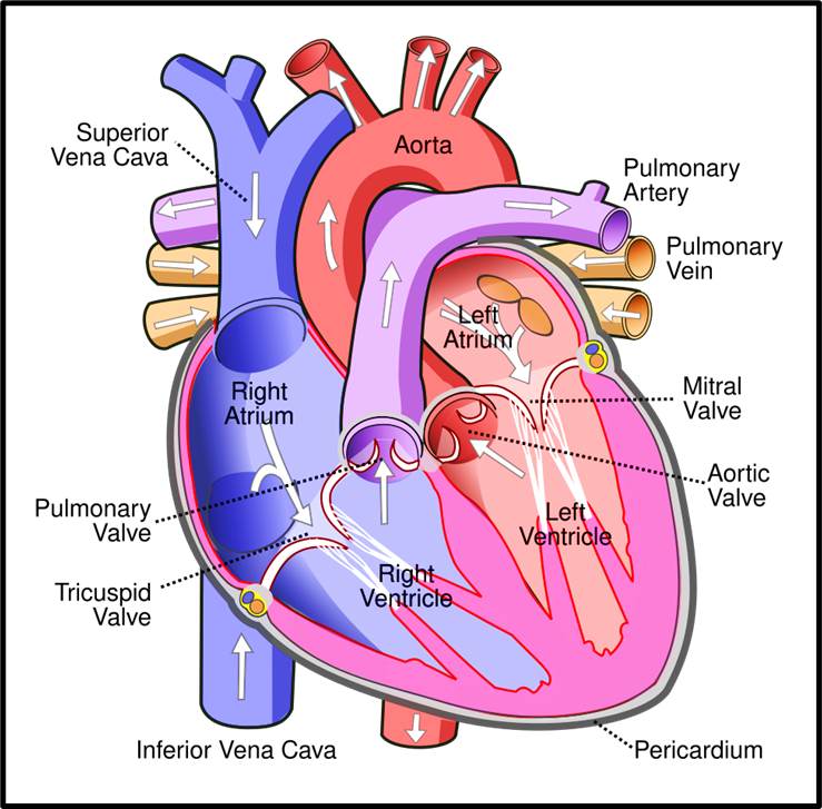
Now structurally, the heart is divided into four chambers:
- Two upper chambers → Atria
- Two lower chambers → Ventricles
The ventricles have thicker muscular walls because they perform the major task of pumping blood to distant organs, while atria mainly receive blood.
Separation within the heart is crucial:
- Interatrial septum → separates right and left atria
- Interventricular septum → separates right and left ventricles
- Atrioventricular septum → separates atria from ventricles
This separation ensures no mixing of oxygenated and deoxygenated blood, which is essential for efficient circulation.
Valves: Ensuring One-Way Traffic
Think of the heart as a system of chambers connected by one-way gates:
- Tricuspid valve → between right atrium and right ventricle
- Bicuspid (Mitral) valve → between left atrium and left ventricle
- Semilunar valves → at the exit of ventricles (pulmonary artery and aorta)
Their role is simple but critical: ➡️ Prevent backflow of blood
This ensures blood flows only in one direction: Atria → Ventricles → Arteries
Cardiac Muscle and Electrical System (Nodal Tissue)
The heart is made of cardiac muscle, which is unique because it is:
- Striated (like skeletal muscle)
- Involuntary (like smooth muscle)
But the real beauty lies in its nodal tissue, which gives the heart its self-excitatory (myogenic) nature.
Key components:
- Sinoatrial node (SAN) → located in right atrium
- Generates impulses at ~70–75 per minute
- Known as the pacemaker of the heart
- Atrioventricular node (AVN) → receives signal from SAN
- AV bundle & Purkinje fibres → spread impulses to ventricles
This coordinated conduction ensures:
➡️ Atria contract first
➡️ Ventricles contract next
Thus, pumping becomes efficient and synchronized.
Function of the Heart (Flow of Blood)
Now let us trace the journey of blood—this is extremely important for conceptual clarity.
Step-by-step flow:
- Oxygenated blood from lungs → Left atrium
- Left atrium → contracts → blood enters Left ventricle
- Left ventricle → pumps blood to entire body
- Deoxygenated blood returns → Right atrium
- Right atrium → contracts → blood enters Right ventricle
- Right ventricle → pumps blood to lungs for oxygenation
So overall:
- Left side → oxygenated blood → body
- Right side → deoxygenated blood → lungs
Cardiac Cycle: One Complete Heartbeat
A cardiac cycle is one complete heartbeat, lasting about 0.8 seconds.
It has following main phases:
- Joint Diastole
All chambers relaxed | Blood flows from atria to ventricles | AV valves open, semilunar valves closed - Atrial Systole
Atria contract (triggered by SAN) | More blood pushed into ventricles - Ventricular Systole
Ventricles contract | AV valves close (prevent backflow) | Semilunar valves open → blood pumped out - Ventricular Diastole
Ventricles relax | Semilunar valves close | AV valves reopen
This cycle repeats continuously, maintaining circulation.
Important Values:
- Heart rate ≈ 72 beats/min
- Stroke volume ≈ 70 mL per beat
- Cardiac output ≈ 5 litres/min
Heart Sounds and ECG
When you listen with a stethoscope:
- “Lub” → closing of tricuspid & bicuspid valves
- “Dub” → closing of semilunar valves
These sounds indicate proper valve functioning.
ECG (Electrocardiograph)
An ECG records the electrical activity of the heart. It shows wave patterns corresponding to different phases of the cardiac cycle and is crucial in diagnosing heart disorders.
Regulation of Cardiac Activity
Even though the heart is myogenic, it is also regulated by the nervous system:
- Sympathetic nerves → increase heart rate and force
- Parasympathetic nerves → decrease heart rate
- Adrenal hormones → enhance cardiac output
So, during exercise → heart rate increases
During rest → heart rate decreases
Types of Circulatory Systems
At the most fundamental level, organisms have evolved two broad types of circulatory systems, depending on how efficiently they need to transport substances like oxygen, nutrients, and waste.
(A) Open Circulatory System
In organisms like arthropods and molluscs, the blood is not strictly confined within vessels. Instead, the heart pumps blood into large vessels, and from there it flows into open spaces called sinuses.
Think of it like an irrigation system where water is released into fields rather than flowing through tightly controlled pipes. Because of this:
- The flow is less regulated
- Pressure is low
- Efficiency is limited
This system works well for organisms with lower metabolic demands.
(B) Closed Circulatory System
In organisms such as annelids and chordates (including humans), blood always flows within a network of vessels—arteries, veins, and capillaries.
Here, the analogy is a well-designed pipeline system:
- Blood flow is precise and controlled
- Pressure can be maintained
- Transport is efficient
This is essential for organisms with higher energy needs, such as vertebrates.
Types of Closed Circulation (Based on Heart Design)
Now, within the closed system, evolution has further refined circulation based on:
- Number of heart chambers
- Number of times blood passes through the heart
(1) Single Circulation
- Found in fish
- Heart has 2 chambers (1 atrium + 1 ventricle)
- Blood passes through the heart once per cycle
Flow pattern: Heart → Gills → Body → Heart
Since blood loses pressure after passing through gills, this system is less efficient.
(2) Incomplete Double Circulation
- Found in amphibians and most reptiles
- Heart has 3 chambers (2 atria + 1 ventricle)
- Blood passes through the heart twice
However, there is a catch:
- Oxygenated and deoxygenated blood partially mix in the ventricle
So, efficiency improves compared to fish, but is still moderate.
(3) Complete Double Circulation
- Found in crocodiles, birds, and mammals (including humans)
- Heart has 4 chambers (2 atria + 2 ventricles)
- Blood passes through the heart twice, but with complete separation
This separation ensures:
- No mixing of oxygenated and deoxygenated blood
- High pressure and efficiency
This is why mammals and birds can sustain high metabolic activity.
Double Circulation in Humans
Humans exhibit complete double circulation, meaning blood travels through two distinct pathways:
(A) Pulmonary Circulation
- Right ventricle → Pulmonary artery → Lungs
- In lungs: blood gets oxygenated
- Returns via pulmonary veins → Left atrium
This circuit is focused on gas exchange (CO₂ removal and O₂ intake).
(B) Systemic Circulation
- Left ventricle → Aorta → Body tissues
- Oxygen and nutrients delivered
- Waste (CO₂, etc.) collected
- Returns via vena cava → Right atrium
This circuit ensures nutrient delivery and waste removal.
Special Circulatory Pathways
- Hepatic Portal System
Blood from the digestive tract first goes to the liver via the hepatic portal vein before entering general circulation.
This allows the liver to → Process nutrients, Detoxify harmful substances - Coronary Circulation
A dedicated network supplies blood directly to the heart muscle (myocardium)—because even the heart needs oxygen to function.
Circulatory System Disorders
Understanding disorders helps you connect theory with real-life implications.
1. Hypertension (High Blood Pressure)
- Blood pressure exceeds 120/80 mm Hg
- Caused by arteriole constriction
- Leads to increased resistance → risk of Artery rupture, Heart disease, Kidney and brain damage
2. Coronary Artery Disease (CAD)
Also called atherosclerosis:
- Arteries supplying the heart get narrowed due to fat, cholesterol, calcium deposits
- Reduces blood flow → risk of heart attack
3. Angina
- Chest pain due to insufficient oxygen supply to heart muscles
- Often a warning sign of underlying coronary issues
4. Heart Failure
- Heart cannot pump enough blood to meet body needs
- Leads to fluid accumulation, especially in lungs
Important distinction:
- Heart attack → damage to heart muscle
- Cardiac arrest → heart stops beating
- Heart failure → reduced pumping efficiency
Blood Pressure: The Driving Force
Blood pressure is the force exerted by blood on vessel walls, and it varies during the cardiac cycle:
- Systolic Pressure: During ventricular contraction
→ Around 120 mm Hg - Diastolic Pressure: During ventricular relaxation
→ Around 80 mm Hg
So, normal BP = 120/80 mm Hg
Arteries experience higher pressure than veins because they receive blood directly from the heart.