
Neural Control and Coordination in Humans
What is the Neural System?
At its core, the neural system is the body’s communication network. It detects changes (stimuli), processes information, and generates appropriate responses.
The basic units of this system are neurons, which are highly specialised cells designed to receive, process, and transmit information.
If we look at evolution:
- In simple organisms like Hydra, the neural system is just a diffused network—no central control.
- In advanced organisms (like humans), it becomes highly organised, with a central command system (brain) and extensive communication lines (nerves).
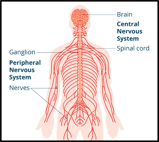
Divisions of the Human Neural System
The human neural system is broadly divided into two major parts:
(A) Central Neural System (CNS)
This is the control centre of the body.
- Composed of the brain and spinal cord
- Responsible for → Processing information, Decision-making, Coordinating responses
Think of it as the “headquarters” where all major decisions are made.
(B) Peripheral Neural System (PNS)
This acts as the communication network.
- Consists of all nerves connecting the body to the CNS
- Functions:
- Carries sensory information to the CNS
- Sends commands from the CNS to muscles and glands
So, if CNS is the headquarters, PNS is the network of wires connecting every part of the body.
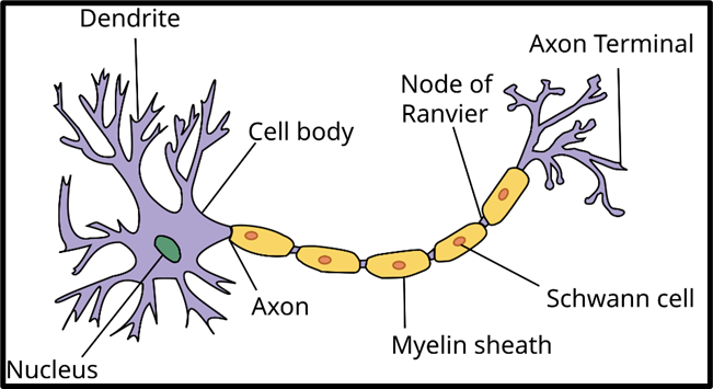
Neuron: Structural and Functional Unit
A neuron is the smallest unit of the neural system, but extremely powerful in function.
Each neuron has three main parts:
(1) Cell Body (Cyton)
- Contains the nucleus and cytoplasm
- Has Nissl’s granules, which are involved in protein synthesis
- Acts as the metabolic centre of the neuron
(2) Dendrites
- Short, branched structures
- Function: Receive signals from other neurons and carry them towards the cell body
(3) Axon
- A long, slender projection
- Function: Transmit signals away from the cell body to other neurons or effector organs (muscles/glands)
Now, axons can be of two types:
- Myelinated axons:
- Covered by a myelin sheath (formed by Schwann cells)
- Myelin is made of lipids and proteins
- Function: Acts as an insulator, increasing the speed of impulse transmission
- Non-myelinated axons:
- No myelin sheath
- Slower transmission
👉 So, myelin sheath is crucial for fast and efficient signal conduction.
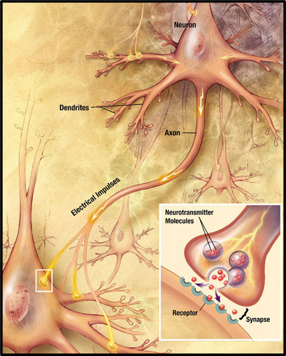
Neural Impulse (How Information Travels)
A neural impulse is essentially an electrical signal.
When a neuron is stimulated:
- There is a rapid change in electrical potential across its membrane
- This generates an action potential
- The action potential travels along the axon like a wave
This is how information moves within the nervous system—electrically along neurons.
Synapse: Junction of Communication
Neurons do not physically touch each other. The gap between them is called a synapse.
A synapse is a junction where one neuron communicates with another neuron or an effector cell.
There are two types:
(1) Electrical Synapse
- Direct connection via gap junctions
- Features:
- Very fast transmission
- Can be bi-directional
- Less common in humans
(2) Chemical Synapse
- Most common type
- Features:
- Uses neurotransmitters (chemical messengers)
- Signal crosses a small gap called the synaptic cleft
- Transmission is:
- Slower than electrical
- Unidirectional (one-way)
👉 So, within a neuron → signal is electrical
👉 Between neurons → signal becomes chemical (in most cases)
Central Neural System (CNS)
Brain: The Master Control Centre
The brain performs a wide spectrum of functions, which can be understood in five broad domains:
| Function Category | Description |
| Voluntary Movements | Enables conscious control over muscle activities, allowing purposeful actions. |
| Involuntary Functions | Regulates automatic processes like heart rate, digestion, and breathing. |
| Homeostasis | Maintains stable internal conditions such as temperature and fluid balance. |
| Circadian Rhythms | Controls biological cycles like sleep-wake patterns and hormonal changes. |
| Higher Functions | Governs thinking, reasoning, emotions, memory, and sensory perception. |
Structural Division of the Brain
The brain is divided into three major regions:
- Forebrain
- Midbrain
- Hindbrain
Let us understand each
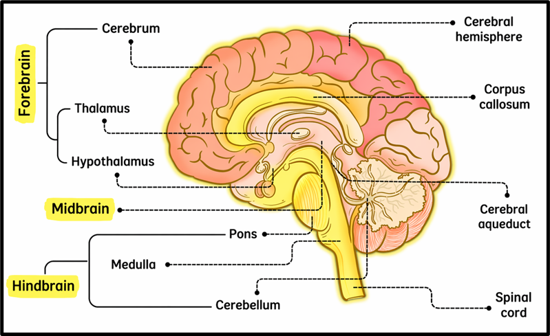
Forebrain: The Centre of Intelligence and Integration
The forebrain is the most developed part in humans and includes → Cerebrum, Thalamus, Hypothalamus, Limbic System
(A) Cerebrum: The Seat of Consciousness
The cerebrum is the largest part of the brain and is responsible for higher mental functions.
- Divided into left and right hemispheres
- These hemispheres are connected by the corpus callosum, enabling coordination between both sides
Internal Organisation
| Component | Location | Composition | Functions | Reason for Colour |
| Cerebral Cortex (Grey Matter) | Outer layer of cerebrum | Neuron cell bodies (cytons) | Processes sensory information, controls motor activity, and higher functions like thinking and reasoning | Appears grey due to dense neuron cell bodies |
| White Matter | Beneath the cerebral cortex | Myelinated axons | Facilitates communication between different regions of the brain | Appears white due to presence of myelin sheath |
(B) Thalamus: The Relay Station
The thalamus acts as a gateway for sensory information:
- Receives sensory signals (except smell)
- Processes and relays them to the cerebral cortex
It ensures that only relevant signals reach higher centres.
(C) Hypothalamus: The Regulatory Hub
The hypothalamus is small but extremely powerful:
- Controls autonomic functions (like temperature, hunger, thirst)
- Links the nervous system with the endocrine system
- Regulates the pituitary gland through hormone release
👉 It is the key structure for maintaining homeostasis.
(D) Limbic System: The Emotional Brain
The limbic system includes structures like:
- Amygdala (emotion processing)
- Hippocampus (memory formation)
Functions:
- Regulates emotions, motivation, and behaviour
- Influences sexual behaviour
- Works closely with the hypothalamus
👉 This is why emotions and memory are deeply interconnected.
Midbrain: The Reflex and Processing Centre
The midbrain connects the forebrain and hindbrain and plays a crucial role in:
- Motor control (especially eye movements)
- Processing visual and auditory stimuli
Key Structures:
Cerebral Aqueduct
- A narrow channel
- Allows flow of cerebrospinal fluid (CSF)
Corpora Quadrigemina
- Responsible for visual and auditory reflexes
- Helps in quick responses like:
- Turning towards sound
- Adjusting eyes to visual stimuli
Hindbrain: The Survival Centre
The hindbrain is essential for basic life-sustaining functions.
It consists of:
(A) Pons
- Acts as a bridge connecting different brain regions
- Contains fibre tracts for signal transmission
(B) Cerebellum
- Highly folded (convoluted) structure → increases surface area and neuron density
Functions:
- Maintains balance and posture
- Coordinates voluntary movements
- Ensures movements are smooth and precise
👉 Without cerebellum, movements become uncoordinated.
(C) Medulla Oblongata
- Connects the brain to the spinal cord
Functions:
- Controls vital autonomic activities → Breathing, Heart rate, Blood pressure, Gastric secretions
👉 This is the life-support centre—damage here can be fatal.
Brain Stem: The Critical Link
The brain stem consists of → Midbrain, Pons, Medulla oblongata
Function:
- Acts as a communication bridge between brain and spinal cord
- Controls basic survival functions
Spinal Cord
The spinal cord is a long, cylindrical structure of nerve tissue extending from the brainstem down through the vertebral column.
It is:
- Protected by vertebrae
- Structurally continuous with the brain
- Functionally essential for linking the brain with the rest of the body
Core Functions of the Spinal Cord
1. Motor Control
- Carries motor signals from the brain to muscles
- Enables voluntary movements like walking, writing, etc.
2. Sensory Perception
- Receives sensory inputs such as → Touch, Temperature, Pain
- Transmits these signals to the brain for interpretation
3. Reflex Actions
- Acts independently of the brain in certain situations
- Enables quick, automatic responses
👉 In essence:
- Ascending pathways → carry sensory signals to the brain
- Descending pathways → carry motor commands to the body
Reflex Action: The Body’s Instant Response System
A reflex action is an automatic, rapid, and involuntary response to a stimulus.
Example:
- Pulling your hand away from a hot object
- Knee-jerk reflex
The key idea is speed and protection—the body responds before conscious thinking.
Mechanism of Reflex Action (Reflex Arc)
Let us follow the sequence logically:
- Sensory Input: A receptor (e.g., skin) detects a stimulus like heat or pain
- Nerve Impulse Transmission: A sensory neuron carries the signal to the spinal cord
- Spinal Cord Processing: The spinal cord processes the information without involving the brain
- Motor Output: A motor neuron sends a signal to the effector (muscle/gland); Immediate response occurs (e.g., withdrawal of hand)
👉 Important insight:
The brain is informed after the action has already occurred. This ensures minimum reaction time, which is critical for survival.
Protection of the Central Neural System (CNS)
Given the importance of the CNS, nature has provided multiple layers of protection.
1. Cranial Meninges: Protective Coverings
The brain and spinal cord are covered by three membranes called meninges:
(A) Dura Mater
- Outer, thick, and tough layer
- Provides mechanical strength
(B) Arachnoid Mater
- Middle, delicate, web-like layer
(C) Pia Mater
- Inner, thin layer closely attached to brain tissue
- Rich in blood vessels, supplying nutrients
👉 Together, these layers act as a protective envelope.
Clinical Link
Meningitis
- It is inflammation of the meninges
- Caused by bacteria, viruses, or fungi
- Can be life-threatening if untreated
2. Skeletal Protection
- The skull (cranium) protects the brain
- The vertebral column protects the spinal cord
3. Cerebrospinal Fluid (CSF): The Protective Cushion
The cerebrospinal fluid (CSF) is a clear, colourless fluid surrounding the brain and spinal cord.
Its functions are extremely important:
| Aspect | Description |
| Protection | Acts as a shock absorber, preventing injury to CNS structures |
| Buoyancy | Reduces effective weight of the brain, preventing compression |
| Chemical Stability | Maintains a stable ionic and chemical environment for neural function |
| Waste Removal | Removes metabolic waste products from the brain |
| Nutrition | Supplies essential nutrients to the brain and spinal cord |
Peripheral Neural System (PNS)
While the CNS is the command centre, the PNS acts as the communication link connecting every part of the body to it.
Types of Nerve Fibres
The nerve fibres of the PNS are of two types:
| Type of Fibre | Direction of Impulse | Function |
| Afferent Fibres | From tissues/organs → CNS | Carry sensory information to the CNS |
| Efferent Fibres | From CNS → tissues/organs | Transmit motor commands to muscles and glands |
Divisions of PNS
| Division | Target/Connection | Type of Control | Function |
| Somatic Neural System | CNS ↔ Skeletal muscles | Voluntary | Controls conscious movements like walking and writing |
| Autonomic Neural System (ANS) | CNS ↔ Internal organs & smooth muscles | Involuntary | Regulates functions like heart rate, digestion, and respiration |
Divisions of Autonomic Nervous System
| Division | Type of Response | Physiological Effects |
| Sympathetic Nervous System | Fight or Flight | Increases heart rate, blood pressure, breathing; dilates pupils; slows digestion |
| Parasympathetic Nervous System | Rest and Digest | Decreases heart rate, blood pressure, breathing; constricts pupils; enhances digestion |
Sensory Reception and Processing
The body continuously interacts with the environment through sensory organs, which detect stimuli and send signals to the CNS for interpretation. These include → Nose, Tongue, Eyes, Ears
1. Nose: Organ of Smell
- Contains olfactory receptors
- These are embedded in mucus
- Connected to the olfactory bulb, which links directly to the limbic system
👉 This explains why smell is strongly linked with memory and emotions
2. Tongue: Organ of Taste
- Contains taste buds with gustatory receptors
- Detect chemicals dissolved in saliva
The brain integrates signals to produce complex flavour perception (taste + smell combined).
3. Eye: Organ of Vision
The human eye is a highly specialised sensory organ designed to detect light and form images.
Structure of the Eye
The eye has three layers:
(A) Outer Layer (Sclera)
- Tough protective layer
- Cornea (transparent part) allows light entry
(B) Middle Layer (Choroid)
- Rich in blood vessels
- Forms:
- Ciliary body → holds lens
- Iris → coloured part controlling light entry
- Pupil → opening that regulates light
(C) Inner Layer (Retina)
- Contains neural cells → Photoreceptors (rods and cones), Bipolar cells, Ganglion cells
Photoreceptors:
- Rods:
- Function: Vision in low light (scotopic vision)
- Contain rhodopsin (Vitamin A-based)
- Cones:
- Function: Colour and daylight vision (photopic)
- Three types: Red, Green, Blue
👉 Combination of cone stimulation → colour perception
👉 Equal stimulation → white light
Special Structures
- Blind Spot → no photoreceptors
- Macula lutea (with fovea) → highest visual acuity (sharpest vision)
Mechanism of Vision
- Light enters through cornea and lens
- Focuses on retina
- Photopigments (opsin + retinal) are activated
- Electrical signals are generated
- Signals travel via optic nerve
- Brain’s visual cortex interprets the image
👉 Vision is not just seeing—it is interpretation by the brain
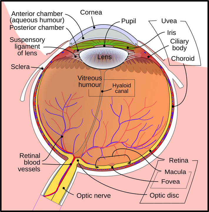
By Rhcastilhos. And Jmarchn, CC BY-SA 3.0, via Wikimedia Commons
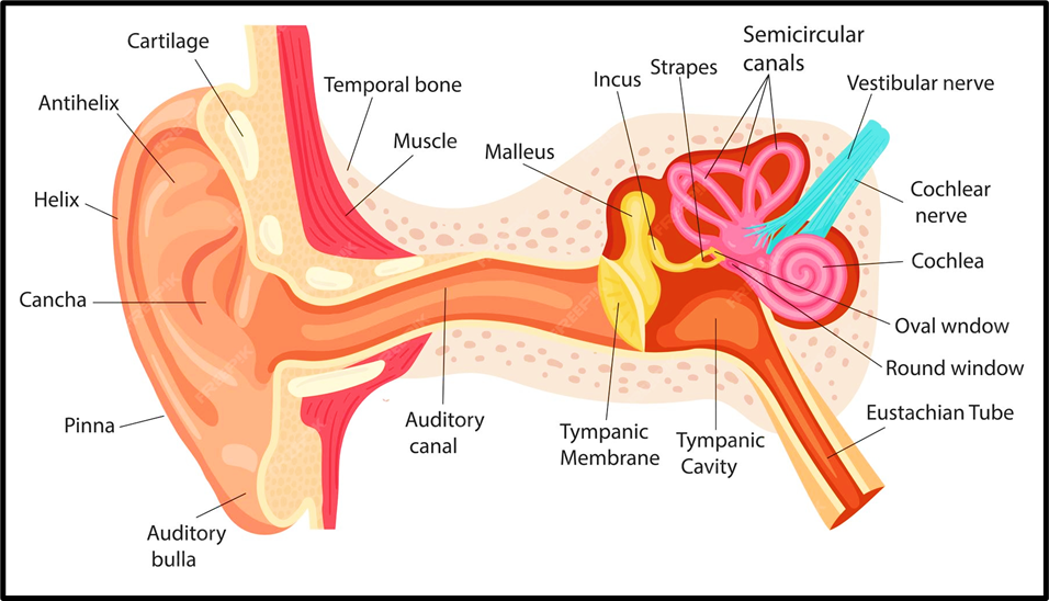
4. Ear: Organ of Hearing and Balance
The ear performs dual functions → Hearing and Maintaining equilibrium
Structure of the Ear
(A) Outer Ear
- Pinna → collects sound
- Auditory canal → directs sound
(B) Middle Ear
- Tympanic membrane (eardrum) → vibrates
- Ossicles (malleus, incus, stapes) → amplify sound
- Eustachian tube → equalises pressure
(C) Inner Ear
Cochlea
- Contains Organ of Corti
- Hair cells act as sound receptors
Vestibular Apparatus
- Maintains balance:
- Semicircular canals → rotational movement
- Utricle & Saccule → linear acceleration and gravity
Mechanism of Hearing
- Sound waves hit the eardrum
- Vibrations pass through ossicles
- Reach cochlea, creating fluid waves
- Hair cells bend and generate impulses
- Signals travel via auditory nerve
- Brain interprets them as sound