
Locomotion and Movement
If you observe life around you, one thing becomes very clear—movement is a defining characteristic of living beings. Even plants move (though slowly), and animals move more visibly.
Now, we must make a subtle but very important distinction:
- Movement → Any change in position of a body part (e.g., blinking, movement of jaw)
- Locomotion → Movement that leads to a change in place or position (e.g., walking, running)
So, all locomotion is movement, but all movement is not locomotion. This conceptual clarity is very important.
Types of Movement in Human Body
At the cellular and tissue level, human body exhibits three types of movement, each based on a different mechanism.
1. Amoeboid Movement
This type of movement is seen in cells like macrophages and white blood cells (WBCs).
- It involves the formation of pseudopodia (false feet)
- The cytoplasm flows in a particular direction (protoplasmic streaming)
- This process is driven by microfilaments (actin filaments)
👉 Think of it like a blob extending itself forward—this is how immune cells chase pathogens.
2. Ciliary Movement
This occurs in organs lined with ciliated epithelium.
- Example: Trachea → Cilia push mucus and dust out
- Example: Female reproductive tract → Cilia help in movement of ovum
👉 Here, movement is not of the whole cell, but of tiny hair-like structures (cilia) that beat rhythmically.
3. Muscular Movement
This is the most familiar type of movement.
- Involves muscle contraction
- Responsible for locomotion and body movements
- Requires coordination of → Muscular system, Skeletal system, Neural system
👉 Without coordination among these three, movement would be impossible.
Muscles: The Engine of Movement
Muscles are specialised tissues of mesodermal origin, forming about 40–50% of body weight.
They have four fundamental properties:
- Excitability → Respond to stimuli
- Contractility → Ability to shorten
- Extensibility → Ability to stretch
- Elasticity → Ability to return to original shape
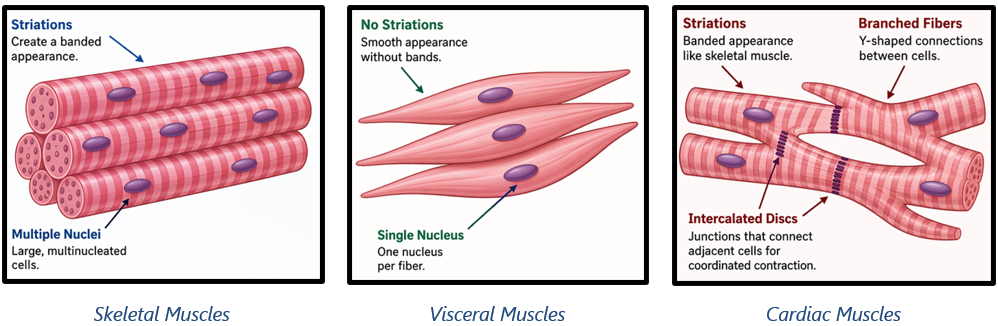
Types of Muscles
| Muscle Type | Location | Appearance | Control Type | Functions |
| Skeletal Muscles | Attached to the skeleton | Striated (striped) | Voluntary | Responsible for body movements and changes in posture |
| Visceral Muscles | Walls of internal organs (e.g., digestive tract, reproductive organs) | Non-striated (smooth) | Involuntary | Help in transport of substances like food and gametes |
| Cardiac Muscles | Heart | Striated (branching) | Involuntary | Pump blood throughout the body |
Structure of Skeletal Muscle: From Macro to Micro
To truly understand movement, we must go deeper into structure.
A skeletal muscle is organized hierarchically:
- Muscle → Fascicles (bundles)
- Fascicle → Muscle fibres (cells)
- Muscle fibre → Myofibrils
- Myofibril → Sarcomeres (functional unit)
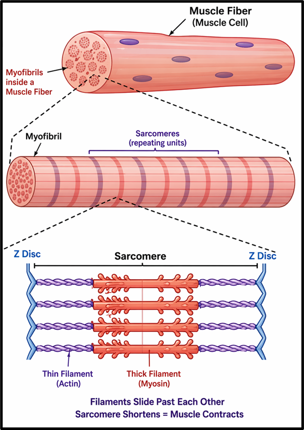
Sarcomere: The Core of Contraction
Each sarcomere contains:
- Actin (thin filaments)
- Myosin (thick filaments)
👉 Their interaction creates the sliding filament mechanism, which leads to contraction.
- Requires:
- Calcium ions (Ca²⁺)
- ATP (Adenosine Triphosphate)
- Produces:
- Dark bands (A bands)
- Light bands (I bands)
Muscle Fibre Types: Functional Specialisation
| Fibre Type | Myoglobin Content | Mitochondria | Metabolism Type | Functional Role |
| Red Fibres | High (gives red colour) | High | Aerobic respiration | Suited for endurance activities |
| White Fibres | Low (gives pale/white colour) | Low | Anaerobic metabolism | Suited for short, intense activities |
Role of Myoglobin
- A red-coloured oxygen-storing pigment in muscles
- Ensures continuous oxygen supply during activity
- Higher in red fibres, hence their colour
Skeletal System: The Structural Foundation
Think of the human body like a building. Muscles provide force, but without a rigid framework, that force would be useless. That framework is the skeletal system.
- Total bones in humans → 206
- Components:
- Bones → Hard, mineralised, provide strength and protection
- Cartilage → Flexible connective tissue, reduces friction and adds flexibility
👉 The skeletal system performs three essential roles:
- Support (gives shape to body)
- Protection (brain, heart, lungs)
- Movement (acts as lever system with muscles)
Division of Skeleton: Axial vs Appendicular
This division is conceptually very important.
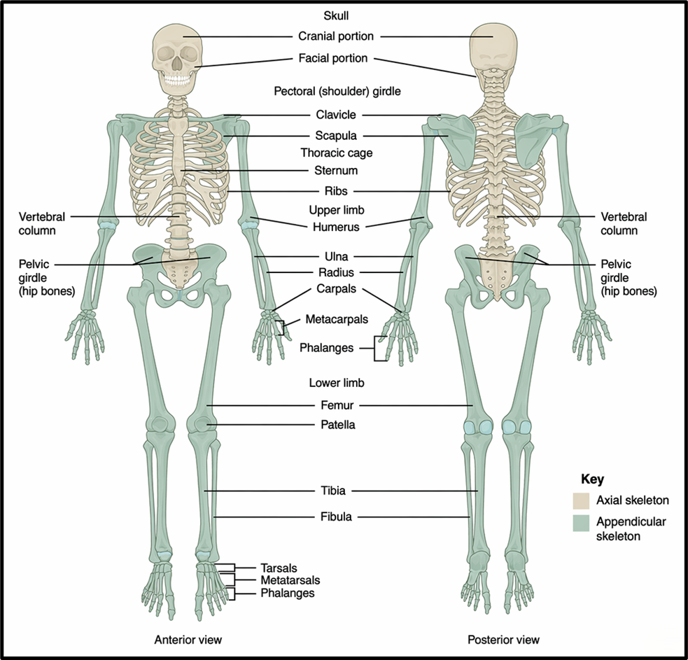
Axial Skeleton (Central Axis of Body)
| Component | Description | Key Details |
| Skull | Protects the brain and forms facial structure | Composed of 22 bones |
| Vertebral Column | Supports the body and protects the spinal cord | Composed of 26 vertebrae: cervical, thoracic, lumbar, sacral, coccygeal |
| Sternum | Flat bone located at the centre of the chest | Forms anterior part of the rib cage |
| Rib Cage | Protects vital organs like heart and lungs | 12 pairs of ribs: 7 true ribs, 3 false ribs, 2 floating ribs |
Rib Cage formed by thoracic vertebrae, ribs, and sternum
- True Ribs: attached to Sternum
- Floating Ribs: not attached to Sternum
- False Ribs: indirectly attached to Sternum
Appendicular Skeleton (Limbs and Girdles)
This part is mainly responsible for movement and locomotion.
1. Limbs
| Sub-Part | Description | Key Details |
| Upper Limbs (Arms) | Enable movement and manipulation | 30 bones: humerus, radius, ulna, carpals, metacarpals, phalanges |
| Lower Limbs (Legs) | Enable locomotion and weight-bearing | 30 bones: femur, tibia, fibula, tarsals, metatarsals, phalanges; patella covers knee |
2. Girdles (Attachment Units)
| Sub-Part | Description | Key Details |
| Pectoral Girdle | Connects upper limbs to axial skeleton | Each side: clavicle and scapula; scapula has glenoid cavity for shoulder joint |
| Pelvic Girdle | Connects lower limbs to axial skeleton | Made of two coxal bones (formed by fusion of ilium, ischium, and pubis) |
👉 Notice the design: upper limbs → more mobility, pelvic girdle → more stability (for weight-bearing)
Joints: The Fulcrum of Movement
Now comes the most interesting mechanical aspect.
A joint is the point where → Two bones meet, or bone meets cartilage
👉 In biomechanics:
- Bone = lever
- Muscle = force
- Joint = fulcrum
Types of Joints
| Joint Type | Structural Feature | Mobility | Example |
| Fibrous Joints | Bones connected by dense fibrous connective tissue | Immovable | Sutures between flat bones of the skull (cranium) |
| Cartilaginous Joints | Bones connected by cartilage | Slightly movable | Joints between vertebrae in the spinal column |
| Synovial Joints | Presence of fluid-filled synovial cavity between bones | Freely movable | Knee, shoulder, hip joints |
Types of Synovial Joints:
- Ball and Socket → Shoulder
- Hinge → Knee
- Pivot → First two vertebrae (neck rotation)
- Gliding → Wrist bones
- Saddle → Base of thumb
👉 This diversity allows the human body to perform highly complex movements.
Disorders of Muscular and Skeletal System
| Disorder | Type/Category | Cause/Mechanism | Key Features/Symptoms |
| Myasthenia Gravis | Autoimmune disorder | Immune system affects neuromuscular junction | Muscle fatigue, weakness, possible paralysis |
| Muscular Dystrophy | Genetic disorder | Progressive degeneration of skeletal muscles | Muscle weakness and wasting over time |
| Tetany | Metabolic condition | Low calcium levels in body fluids | Rapid, involuntary muscle spasms |
| Arthritis | Inflammatory condition | Inflammation of joints | Joint pain, swelling, stiffness |
| Osteoporosis | Degenerative condition | Decreased bone mass (often linked to reduced estrogen levels) | Increased fracture risk, brittle bones |
| Gout | Metabolic disorder | Deposition of uric acid crystals in joints | Severe joint inflammation, pain |