Heredity
Imagine a child who has their father’s eyes and their mother’s smile. You’ve seen this countless times — and perhaps wondered:
How exactly does a child inherit traits from their parents?
Why do siblings from the same parents sometimes look completely different from each other?
And here’s the big question that has puzzled humanity for centuries — where did all the diverse life forms on Earth come from?
These two seemingly different questions — How are traits inherited? and How did life evolve? — are in fact deeply connected.
💡 Think of heredity as the ‘rule book’ of biological inheritance — the instructions passed from one generation to the next. Evolution, on the other hand, is what happens when those rules get ‘edited’ over millions of years. Together, they explain the entire diversity of life on Earth.
The primary outcome of any reproduction — whether in bacteria or in humans — is the creation of new individuals. But here’s what’s remarkable: these new individuals don’t look completely random. They resemble their parents. This phenomenon is explained by Heredity.
Heredity refers to the rules and mechanisms by which traits are transmitted from parents to offspring. Inheritance is the actual process of passing those traits down. Think of heredity as the ‘subject’ and inheritance as the ‘process.’
Key Concepts at a Glance
- Inherited Traits: Characteristics passed from parents to offspring — e.g., eye colour, blood type, height, hair texture. These are governed by genes and tend to be consistent within families.
- Variations: Differences within a species that arise despite having a common genetic framework. Variations occur due to (a) genetic mutations, (b) gene recombination during sexual reproduction, and (c) environmental influences. These variations are crucial — without them, evolution cannot occur!
💡 Here’s an analogy: Heredity is like a photocopier — it tries to replicate the original. Variation is like a photocopier that occasionally makes errors. These ‘errors’ in nature are not bugs — they are features that drive evolution!
Principles of Inheritance and Variation
Mendel’s Experiments on Inheritance
Before Gregor Mendel, people believed in ‘blending inheritance’ — the idea that parental traits simply merge in offspring like mixing paints. Mendel’s experiments shattered this myth and gave us the first scientific basis for inheritance. This is why he is rightly called the ‘Father of Genetics.’
Why Did Mendel Choose Pea Plants?
Not every scientist chooses their experimental subject wisely, but Mendel did. His choice of the garden pea plant (Pisum sativum) was brilliant for the following reasons:
- Pea plants are easy to grow, maintain, and study over multiple generations.
- They are naturally self-pollinating, but can also be cross-pollinated artificially — giving the experimenter full control.
- Being annual plants, many generations could be studied within a short time frame.
- They exhibit several distinct, contrasting traits (e.g., tall vs. dwarf, round vs. wrinkled seeds) — making observations clear-cut.
Mendel’s Method: Controlled Cross-Pollination
Mendel focused on seven pairs of contrasting traits and used artificial (controlled) cross-pollination to study how each trait is inherited. He began with 14 true-breeding (purebred) varieties — plants that consistently show the same trait over generations through self-pollination.
💡 A ‘true-breeding line’ is like a purebred dog breed — it always produces offspring with the same traits when mated with its own kind.
The Seven Traits Mendel Studied
| Trait | Dominant Form | Recessive Form |
| Stem height | Tall | Dwarf (short) |
| Seed shape | Round | Wrinkled |
| Seed colour | Yellow | Green |
| Pod shape | Inflated (smooth) | Constricted |
| Pod colour | Green | Yellow |
| Flower colour | Purple | White |
| Flower position | Axial (along stem) | Terminal (at tip) |
Monohybrid Cross — Inheritance of One Gene
Mendel crossed true-breeding tall plants (TT) with true-breeding dwarf plants (tt). The result? All plants in the First Filial generation (F₁) were tall. The dwarf trait seemed to disappear! But when these F₁ plants (Tt) were allowed to self-pollinate, the Second Filial generation (F₂) produced both tall and dwarf plants in a 3:1 ratio (3 tall : 1 dwarf).
💡 The dwarf trait didn’t disappear — it was hiding! Mendel realised that traits come in two versions (alleles), and one can ‘mask’ the other. The hidden trait reappears when two copies of it come together in F₂.
Key Observations from Monohybrid Cross
- No Blending of Traits: F₁ plants showed only one parent’s trait. Traits do not blend — they remain distinct.
- Genes and Alleles: Traits are governed by ‘factors’ (now called genes). Each gene has two versions called alleles. For height: ‘T’ (tall, dominant) and ‘t’ (dwarf, recessive).
- Genotype vs Phenotype:
- Genotype = genetic makeup (TT, Tt, tt).
- Phenotype = visible trait (tall or dwarf).
- Both TT and Tt plants are tall in phenotype, but different in genotype.
- Dominant and Recessive Traits: In the presence of ‘T’, the plant is tall — even if only one copy of T is present. Dwarf (tt) appears only when both alleles are recessive.
- Segregation of Alleles: Alleles separate during gamete formation. Each gamete carries only ONE allele.
- F₂ genotypic ratio = 1TT : 2Tt : 1tt → Phenotypic ratio = 3 tall : 1 dwarf.
Dihybrid Cross — Inheritance of Two Genes
When Mendel crossed plants differing in two traits simultaneously (e.g., seed colour — yellow/green, and seed shape — round/wrinkled):
- P generation: Yellow-Round (YYRR) × Green-Wrinkled (yyrr)
- F₁ generation: All offspring were Yellow-Round (YyRr) — both dominant traits expressed.
- F₂ generation: 4 phenotypic classes appeared in a 9:3:3:1 ratio — 9 Yellow-Round : 3 Yellow-Wrinkled : 3 Green-Round : 1 Green-Wrinkled.
This spectacular result led Mendel to his third law — the Law of Independent Assortment.
Mendel’s Laws of Inheritance
Based on his meticulous experiments, Mendel formulated three fundamental laws — the bedrock of classical genetics:
1. Law of Dominance
In a pair of alleles, one allele (dominant) masks the expression of the other (recessive). In a heterozygous organism (Tt), only the dominant trait is visible. The recessive trait only appears in the homozygous recessive condition (tt).
2. Law of Segregation
Each organism carries two alleles for every trait. During the formation of gametes (reproductive cells), these two alleles separate (segregate) from each other. Each gamete receives only ONE allele. When fertilisation occurs, the offspring inherits one allele from each parent — restoring the pair.
Example:
- A Tt plant produces gametes carrying either T or t. During self-pollination → F₂ ratio: 1TT : 2Tt : 1tt → Phenotypic ratio: 3 tall : 1 dwarf.
3. Law of Independent Assortment
Genes for different traits are inherited independently of each other — one trait’s inheritance does NOT influence another’s. This holds true when genes are on different chromosomes (or far apart on the same chromosome).
Example: In the dihybrid cross, seed colour genes and seed shape genes were inherited independently, giving the 9:3:3:1 ratio.
💡 Quick recap of terms:
• Gene = a section of DNA coding for a specific trait.
• Allele = alternative forms of the same gene (e.g., ‘T’ and ‘t’).
• Heterozygous = two different alleles (Tt).
• Homozygous Dominant = two dominant alleles (TT).
• Homozygous Recessive = two recessive alleles (tt).
Beyond Mendel — Exceptions and Extensions
Incomplete Dominance
Mendel’s law of dominance assumes one allele completely dominates the other. But what if neither is fully dominant? This is called Incomplete Dominance — where the heterozygous phenotype is a ‘blend’ or intermediate of both parental traits.
Classic Example — Snapdragon Flowers: Red (RR) × White (rr) → F₁ = all PINK (Rr). The pink colour is intermediate — neither parent’s colour dominates completely. F₂ = 1 Red : 2 Pink : 1 White.
💡 Here the genotypic and phenotypic ratios in F₂ are BOTH 1:2:1 — unlike Mendelian crosses where phenotypic ratio is 3:1. This is a classic exam trick!
Co-dominance
In co-dominance, both alleles in a heterozygous organism are fully and simultaneously expressed — neither is recessive. The classic example is the ABO blood group system in humans.
| Genotype (Genetic Makeup) | Phenotype (Blood Group) |
| IAIA or IAi | Blood Group A |
| IBIB or IBi | Blood Group B |
| IAIB | Blood Group AB — both A and B antigens expressed (Co-dominance) |
| ii | Blood Group O — no antigens expressed |
The gene ‘I’ has three alleles: IA, IB, and i. This is also an example of multiple allelism — more than two alleles for a single gene in the population (though each individual carries only two).
Chromosomal Theory of Inheritance
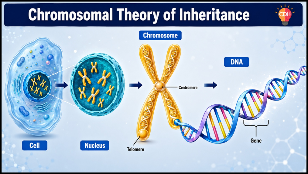
Mendel didn’t know about chromosomes — they were discovered later. In the early 1900s, Walter Sutton and Theodore Boveri independently observed that the behaviour of chromosomes during meiosis perfectly parallels Mendel’s laws. This led to the Chromosomal Theory of Inheritance.
Key Points of the Theory
- Genes are located on chromosomes — at specific positions called ‘loci.’
- Chromosomes come in homologous pairs (like Mendel’s paired alleles).
- During meiosis, homologous chromosomes separate → explains Mendel’s Law of Segregation.
- Random alignment of chromosome pairs during meiosis → explains the Law of Independent Assortment.
| Feature | Genes | Chromosomes |
| Definition | Segments of DNA coding for proteins or RNA | Thread-like structures of DNA + proteins |
| Size | Smallest functional unit of heredity | Larger structures composed of many genes |
| Structure | Linear sequence of nucleotides | DNA coiled around histone proteins |
| Number in Humans | ~20,000–25,000 genes | 46 chromosomes (23 pairs) |
| Location | On chromosomes | Nucleus of eukaryotic cells |
| Function | Encode proteins, determine traits | Carry and transmit genetic information |
Thomas Hunt Morgan’s Experiments — Sex-Linked Inheritance
Working with Drosophila melanogaster (fruit flies), Thomas Hunt Morgan provided the experimental evidence for the chromosomal theory. His most significant contribution was the concept of sex-linked inheritance — the inheritance of genes located on sex chromosomes (X and Y).
Sex Chromosomes at a Glance
- Females: XX — two X chromosomes
- Males: XY — one X and one Y chromosome
X-Linked Inheritance
Since the X chromosome carries more genes than the Y chromosome, genes on the X chromosome follow a distinct pattern. Males (XY) are particularly vulnerable to X-linked recessive traits — because they have only ONE X chromosome, a single recessive allele is enough to express the trait.
- Examples: Haemophilia, Red-Green Colour Blindness, Duchenne Muscular Dystrophy.
- If a mother is a carrier, her sons have a 50% chance of being affected.
- Daughters can only be affected if BOTH parents pass the recessive allele.
Y-Linked Inheritance
Genes exclusively on the Y chromosome pass directly from father to all sons — never to daughters (who don’t have a Y chromosome). This is less common as Y carries fewer genes. Example: certain types of male infertility.
Linkage and Recombination
When Mendel discovered independent assortment, he was lucky — his chosen traits happened to be on different chromosomes. But what about genes that sit on the same chromosome? They tend to be inherited together — this is called Linkage.
- Complete Linkage: Genes very close on the same chromosome — always inherited together, no recombination.
- Incomplete Linkage: Genes somewhat close — can occasionally be separated by ‘crossing over,’ leading to recombination.
Recombination is the exchange of genetic material between homologous chromosomes during meiosis via crossing over. It creates new combinations of alleles not present in either parent — a major source of genetic variation.
💡 Crossing over: Think of two chromosome ‘ladders’ lying side by side — they swap some rungs. The result? New ‘hybrid’ ladders that carry a mix of both parents’ genetic information. This is why siblings can look different from each other even with the same parents.
Genetic Mapping
Alfred Sturtevant, Morgan’s student, created the first genetic map — a diagram showing the positions of genes on a chromosome. The distance between genes is measured in map units (cM, centimorgans). 1 cM = 1% recombination frequency between two genes. The greater the distance, the higher the recombination frequency.
Polygenic Inheritance
Most of Mendel’s traits had just two clear options — tall or dwarf, purple or white. But many real-world traits show a continuous range of phenotypes. This is because they are controlled by multiple genes acting together — this is Polygenic Inheritance.
Example: Human skin colour is controlled by multiple genes. Each dominant allele (say A, B, or C) adds a bit of darkness, while each recessive allele (a, b, c) adds lightness. A person with AABBCC has the darkest skin; aabbcc has the lightest. The spectrum in between represents all the intermediate tones we observe.
- Other polygenic traits in humans: Height, eye colour, weight, intelligence, behaviour, and susceptibility to certain diseases.
- Environmental influence: Environmental factors (like sun exposure) can also modify polygenic traits, making them even more variable.
💡 Polygenic inheritance is ADDITIVE — each gene contributes a small, cumulative effect to the overall phenotype. This is why these traits show a bell-curve distribution in a population.
Pleiotropy — One Gene, Many Effects
The reverse of polygenic inheritance! In Pleiotropy, a single gene affects multiple traits or characteristics simultaneously. This happens because the gene influences metabolic pathways that have wide-ranging effects.
- Example: Phenylketonuria (PKU) — a mutation in one gene causes both intellectual disability AND abnormal hair and skin pigmentation. One gene, multiple consequences.
Sex Determination — Who Decides the Sex?
A timeless question — is the sex of a child determined by the mother or the father? Science gives us a clear, unambiguous answer. The sex is determined by specific sex chromosomes, and in humans, it is the father’s sperm that decides the sex of the child (as we’ll see below). Let’s explore the different sex determination systems in the animal kingdom.
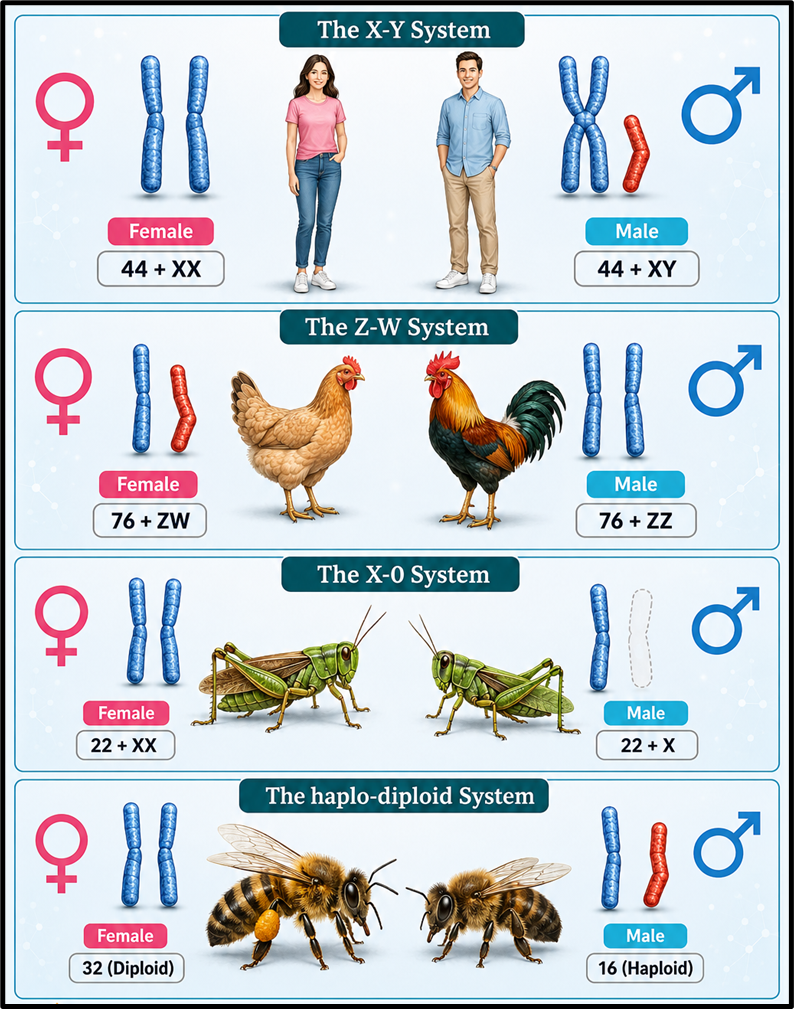
Sex Determination Systems
| System | Found In | Females | Males | Who Determines Sex? |
| XO System | Some insects (e.g., grasshoppers) | XX | XO (only one X) | Male (sperm carries X or O) |
| XY System | Humans, most mammals, some insects | XX | XY | Male (sperm carries X or Y) |
| ZW System | Birds, some reptiles & fish | ZW | ZZ | Female (egg carries Z or W) |
| Haplodiploid System | Honeybees, ants, wasps | Diploid (from fertilised eggs) | Haploid (from unfertilised eggs) | Determined by ploidy level |
Environmental Sex Determination
Not all organisms use chromosomes for sex determination. In many turtles, crocodiles, and certain fish, the temperature during egg incubation determines the sex of offspring.
- Eggs incubated at lower temperatures (22–27°C) → produce Males
- Eggs incubated at higher temperatures (30°C and above) → produce Females
This has critical implications for wildlife conservation — climate change is skewing sex ratios in many reptile populations!
Sex Determination in Humans — The XY System
Humans have 23 pairs of chromosomes: 22 pairs are autosomes (same in both sexes) and 1 pair are sex chromosomes. Females are XX and males are XY.
How it works: Males produce two types of sperm — 50% carry X, 50% carry Y. Females produce only X-carrying eggs.
- X sperm + X egg → XX → Girl
- Y sperm + X egg → XY → Boy
💡 In humans, it is ALWAYS the father’s sperm that determines the sex of the child — NOT the mother’s egg. This is scientifically established and has immense social implications in the Indian context where mothers are blamed for the sex of children!
📌 Male heterogamety = Males produce different types of gametes (X and Y) → XO and XY systems.
📌 Female heterogamety = Females produce different types of gametes (Z and W) → ZW system.
Mutation — When the Blueprint Changes
A mutation is any change in the DNA sequence of an organism. Mutations alter the genotype and may or may not affect the phenotype. They are a primary source of genetic variation — and therefore, a driver of evolution.
Types of Mutations
- Chromosomal Mutations: Involve deletion, insertion, or duplication of large DNA segments → can cause cancer and severe genetic disorders.
- Point Mutations: A change in a single base pair of DNA. Example: Sickle-cell anaemia is caused by just ONE base change.
- Frame-Shift Mutations: Insertion or deletion of base pairs that shifts the entire reading frame of the genetic code, usually causing severe disruption.
- Mutagens: Agents that cause mutations. These include chemical substances (e.g., certain dyes, benzene) and physical agents like UV radiation.
Genetic Disorders
When mutations or chromosomal abnormalities disrupt normal biological functioning, we get genetic disorders. These are broadly classified into two categories:
- Mendelian Disorders: Caused by mutations in a single gene. Follow Mendel’s laws of inheritance.
- Chromosomal Disorders: Caused by changes in the number or structure of chromosomes.
Mendelian Disorders
These follow predictable inheritance patterns — autosomal or sex-linked, dominant or recessive. Pedigree analysis (family tree diagrams) helps trace their inheritance across generations.
Colour Blindness
Colour blindness is a sex-linked recessive disorder caused by mutations in genes on the X chromosome. It affects the photoreceptor cells (cones) in the retina. It is more common in males (~8%) than females (~0.4%) because males have only one X chromosome.
| Type | What’s Affected | Subtype |
| Red-Green Colour Blindness | Difficulty distinguishing red and green | Protanopia (red), Deuteranopia (green) |
| Blue-Yellow Colour Blindness | Difficulty distinguishing blue and yellow | Tritanopia (blue) |
| Complete Colour Blindness (Monochromacy) | Cannot perceive any colour | Only shades of grey are visible |
Haemophilia
Haemophilia is an X-linked recessive disorder where blood clotting is impaired due to deficiency of clotting proteins, causing excessive bleeding even from minor injuries.
- Males are primarily affected (only one X chromosome — no backup copy).
- Females with two X chromosomes would need BOTH to carry the mutation — extremely rare.
- Historical note: Queen Victoria was a carrier, spreading haemophilia across the royal families of Europe.
Sickle-Cell Disease
Sickle-Cell Disease is an autosomal recessive disorder affecting haemoglobin — the oxygen-carrying protein in Red Blood Cells (RBCs). A point mutation causes RBCs to become crescent (sickle) shaped instead of the normal flexible disc shape.
- Consequence: Sickle-shaped RBCs are rigid, can block blood vessels, and don’t carry oxygen efficiently.
- Symptoms: Pain crises, anaemia, jaundice, stroke, organ failure.
- Treatment: Blood/bone marrow transplant (only cure); Hydroxyurea (oral medication to manage symptoms); Gene therapy showing promising results.
India and Sickle-Cell Anaemia
India has the second-highest burden of sickle-cell anaemia globally (after African countries). It is most prevalent among tribal populations and in 15 Indian states, with Maharashtra leading in incidence.
| Initiative | Key Detail |
| National Health Mission (NHM) | Supports state governments in prevention and management of sickle-cell anaemia. |
| State Haemoglobinopathy Mission | Established in Madhya Pradesh (launched 2021) for screening and managing sickle-cell anaemia. |
| National Sickle Cell Anaemia Elimination Mission (NSCAEM) | Introduced in Union Budget 2023. Aims to eliminate it as a public health problem by 2047. Covers 17 states. |
Phenylketonuria (PKU)
An autosomal recessive metabolic disorder where the enzyme to convert phenylalanine into tyrosine is absent. Phenylalanine accumulates and forms phenylpyruvic acid, leading to intellectual disability and abnormal pigmentation. This is also an example of Pleiotropy — one gene, multiple effects.
Thalassemia
Thalassemia is an autosomal recessive inherited blood disorder where the body produces insufficient healthy haemoglobin, resulting in anaemia.
| Feature | Thalassemia Major (Cooley’s Anaemia) | Thalassemia Minor (Carrier State) |
| Severity | Severe | Mild |
| Genetics | Homozygous — defective genes from both parents | Heterozygous — defective gene from one parent |
| Symptoms | Severe anaemia, bone deformities, fatigue, jaundice, growth issues | Mild anaemia, often asymptomatic |
| Treatment | Regular blood transfusions; possible bone marrow transplant | Usually no treatment needed; occasional iron supplements |
| Impact on Life | Significant — reduced lifespan without treatment | Minimal to no impact; normal lifespan |
📌 UPSC Connect: India has the LARGEST number of children with Thalassemia Major in the world. Gene therapy is being explored as a future cure.
Chromosomal Disorders
These arise from abnormalities in the number or structure of chromosomes — often due to errors in cell division (meiosis). The key mechanism is non-disjunction — failure of chromosomes to separate properly.
- Aneuploidy: Gain or loss of ONE or more chromosomes (e.g., trisomy, monosomy).
- Polyploidy: Gain of an ENTIRE extra set of chromosomes. Common in plants; lethal in animals.
Down Syndrome (Trisomy 21)
Caused by an extra copy of chromosome 21, giving a total of 47 chromosomes. First described by Langdon Down.
| Type | Description | Frequency |
| Trisomy 21 | All cells have three separate copies of chromosome 21 | ~95% of Down Syndrome cases |
| Mosaic Down Syndrome | SOME cells have trisomy 21, others have normal 46 | ~2% of cases |
| Translocation Down Syndrome | Extra chr. 21 attached to another chromosome | ~3% of cases |
- Physical Features: Flattened face, almond-shaped eyes, short neck, small ears, single palmar crease, low muscle tone.
- Cognitive Issues: Mild to moderate intellectual disability, delayed speech.
- Health Risks: Congenital heart defects (~50%), thyroid disorders, risk of Alzheimer’s disease.
Klinefelter’s Syndrome (47, XXY)
Affects males who are born with an extra X chromosome — making them 47, XXY instead of the usual 46, XY. Caused by non-disjunction during parental cell division.
- Physical: Taller stature, longer limbs, reduced muscle mass, enlarged breast tissue (gynecomastia), small testicles.
- Reproductive: Infertility due to impaired sperm production; low testosterone levels.
- Cognitive: Delayed speech, learning difficulties, attention issues.
- Health Risks: Osteoporosis, type 2 diabetes, cardiovascular disease, increased risk of breast cancer.
Turner’s Syndrome (45, XO)
Affects females who are missing one X chromosome — giving them only 45 chromosomes (45, XO) instead of the usual 46, XX. Caused by non-disjunction.
- Physical: Short stature, broad chest with widely spaced nipples, webbed neck, low hairline, swelling of hands and feet.
- Reproductive: Underdeveloped ovaries, infertility, absent or delayed menstrual cycles.
- Health Risks: Heart defects, kidney abnormalities, osteoporosis (low estrogen), hypothyroidism.
Molecular Basis of Inheritance
So far, we’ve spoken about ‘genes’ and ‘alleles’ abstractly. Now let’s go deeper — to the molecular level — to understand what genes actually ARE, how genetic information is stored, and how it flows within a cell. This part explores the central dogma of molecular biology.
Nucleic Acids — The Information Molecules
Genes are made of Nucleic Acids — naturally occurring chemical compounds that carry genetic information. There are two types:
- DNA (Deoxyribonucleic Acid): The primary genetic material in almost all living organisms. Long-term storage of genetic information.
- RNA (Ribonucleic Acid): Works as an intermediate — carries information from DNA and helps translate it into proteins. Also serves as the genetic material in some viruses (retroviruses).
DNA — The Double Helix
DNA is a double-stranded long polymer made of smaller units called nucleotides. It carries the complete genetic blueprint of an organism. DNA was first identified as an acidic substance in the cell nucleus by Friedrich Meischer in 1869 (he called it ‘Nuclein’). Its famous double helix structure was proposed by James Watson and Francis Crick in 1953.
Structure of a Nucleotide
Each nucleotide — the building block of DNA (and RNA) — has THREE components:
- Phosphate group: Contains phosphorus; gives DNA its acidic nature.
- Pentose sugar: Deoxyribose in DNA; Ribose in RNA.
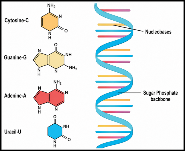
- Nitrogenous base: The information-carrying part. Four types in DNA: Adenine (A), Guanine (G), Cytosine (C), Thymine (T). In RNA, Thymine is replaced by Uracil (U).
| Type | Nitrogenous Bases | Structure |
| Purines | Adenine (A) and Guanine (G) | Larger, double-ring structure |
| Pyrimidines | Cytosine (C), Thymine (T) — in DNA; Uracil (U) — in RNA | Smaller, single-ring structure |
The Double Helix Structure
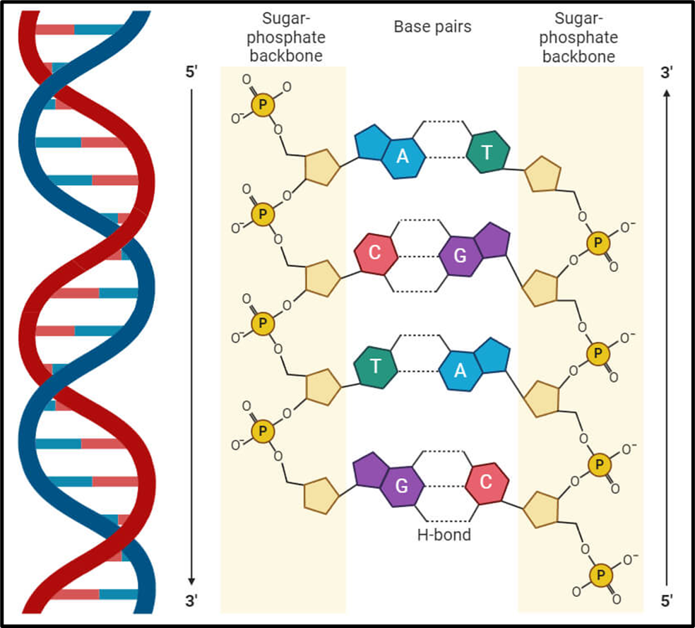
Two polynucleotide strands coil around each other forming a double helix — like a twisted ladder. The sugar-phosphate backbone forms the ‘rails’ of the ladder, and the nitrogenous bases form the ‘rungs.’ The two strands run anti-parallel (opposite directions).
Base Pairing Rules (Chargaff’s Rules)
- Adenine (A) pairs with Thymine (T) — connected by 2 hydrogen bonds
- Guanine (G) pairs with Cytosine (C) — connected by 3 hydrogen bonds
💡 The G≡C bond (3 H-bonds) is STRONGER than the A=T bond (2 H-bonds). This is why DNA regions rich in G-C are more thermally stable — they require more energy to ‘unzip.’
This specific, complementary base pairing is crucial: if the sequence of one strand is known, the other can be predicted entirely. It is also the molecular basis of DNA replication.
Packaging of DNA in Cells
The human genome contains about 3 billion base pairs of DNA. If stretched out, a single cell’s DNA would be about 2 metres long! Yet it fits inside a nucleus about 6 micrometres in diameter. How? Through elegant packaging:
- In Prokaryotes (e.g., E. coli): No defined nucleus. DNA is compacted into a region called the nucleoid, held by positively charged proteins that neutralise DNA’s negative charge.
- In Eukaryotes: DNA wraps around histone proteins → forming nucleosomes (like ‘beads on a string’). Nucleosomes coil further → chromatin fibres → chromosomes during cell division.
| Feature | Chromatin | Chromosomes |
| Structure | Less condensed DNA wrapped around histones | Highly condensed chromatin with two sister chromatids |
| Function | DNA replication, transcription, repair | Accurate distribution of DNA during cell division |
| Appearance | ‘Beads on a string’ | Thread-like structures |
| Presence | During interphase (non-dividing cells) | During cell division (mitosis/meiosis) |
RNA — The Messenger and More
RNA is a mostly single-stranded polymer of ribonucleotides. It is found in both the nucleus and cytoplasm. In some viruses, RNA itself serves as the genetic material (e.g., HIV, coronavirus) — such viruses are called retroviruses.
Types of RNA
| Type of RNA | Full Name | Function | Abundance |
| mRNA | Messenger RNA | Carries genetic information from DNA to ribosomes; serves as template for protein synthesis | ~3–5% of total cell RNA |
| rRNA | Ribosomal RNA | Combines with proteins to form ribosomes; forms peptide bonds during translation | ~80% of total cell RNA |
| tRNA | Transfer RNA | Acts as adaptor — reads mRNA codons and brings the correct amino acid to the ribosome | ~15% of total cell RNA |
| miRNA | MicroRNA | Short, non-coding RNA that silences gene expression at post-transcriptional level | Variable |
| siRNA | Small Interfering RNA | Non-coding RNA involved in gene regulation; being studied as therapeutic agent | Variable |
DNA vs RNA — A Comparative View
| Feature | DNA | RNA |
| Sugar | Deoxyribose | Ribose |
| Strands | Double-stranded | Single-stranded (usually) |
| Nitrogenous Bases | A, T, G, C | A, U, G, C (Uracil instead of Thymine) |
| Location | Mainly in nucleus | Nucleus and cytoplasm |
| Length | Longer (contains many genes) | Shorter (corresponds to a gene/function) |
| Types | One main type | Multiple types (mRNA, tRNA, rRNA, miRNA, siRNA) |
| Genetic Material? | Yes — in almost all organisms | Yes — only in some viruses (retroviruses) |
| Stability | More stable, less reactive | Less stable, more reactive |
| Mutation Rate | Lower (DNA polymerase has proofreading) | Higher (RNA polymerase lacks proofreading) |
Genetic Code — The Language of Genes
How does a sequence of four DNA bases (A, T, G, C) code for 20 different amino acids that make up all proteins? The answer lies in the Genetic Code — the set of rules by which nucleotide sequences are translated into amino acid sequences.
Key Features of the Genetic Code
- Triplet Codon: Each amino acid is coded by a group of 3 nucleotides called a CODON. (4³ = 64 possible codons for 20 amino acids)
- Total Codons: 64 codons — 61 code for amino acids, 3 are STOP codons (no amino acid).
- Degenerate (Redundant): Multiple codons can code for the same amino acid. E.g., UCU, UCC, UCA, UCG all code for Serine.
- Universal: Nearly identical across ALL living organisms — from bacteria to humans. This is powerful evidence of a common evolutionary origin!
- Non-overlapping: Codons are read sequentially, without any overlap.
- Start Codon: AUG — codes for methionine; initiates protein synthesis.
- Stop Codons: UAA, UAG, UGA — signal the end of translation. They don’t code for any amino acid.
Process of Protein Synthesis — The Central Dogma
💡 The Central Dogma of Molecular Biology: DNA → RNA → Protein. Genetic information flows in one direction. DNA is transcribed into RNA, and RNA is translated into protein.
Step 1: DNA Replication
Before a cell divides, its DNA must be faithfully copied — this is DNA Replication. It is semiconservative — each new DNA molecule has one original (parental) strand and one newly made strand.
| Enzyme | Function |
| Helicase | Unwinds the DNA double helix into two single strands |
| DNA Gyrase | Reduces the supercoiling that forms as DNA unwinds |
| Primase | Creates short RNA primers to initiate DNA synthesis |
| DNA Polymerase III | Main enzyme — adds nucleotides to build new DNA strands |
| DNA Polymerase I | Fills small DNA gaps during replication and repair |
| DNA Polymerase II | Proofreads and corrects errors |
| DNA Ligase | Joins discontinuously synthesised DNA fragments (Okazaki fragments) |
Step 2: Transcription — DNA to RNA
Transcription is the process of copying the genetic information from DNA into messenger RNA (mRNA). The key enzyme is RNA polymerase. In eukaryotes, this occurs in the NUCLEUS; the mRNA then travels to the cytoplasm.
- Initiation: RNA polymerase binds to the promoter region of DNA. DNA unwinds, exposing the template strand.
- Elongation: RNA polymerase moves along DNA, synthesising complementary RNA by adding ribonucleotides. (T in DNA → A in RNA; A in DNA → U in RNA)
- Termination: RNA polymerase reaches the terminator sequence and releases the newly made mRNA strand.
Step 3: Translation — mRNA to Protein
Translation is the process by which the genetic code in mRNA is ‘read’ by ribosomes to build a specific protein. tRNA molecules bring amino acids corresponding to each mRNA codon.
- Initiation: Ribosome binds to mRNA at the Start Codon (AUG). Initiator tRNA carrying methionine binds to AUG.
- Elongation: Ribosome moves along mRNA. Each codon is matched by a tRNA anticodon carrying the correct amino acid. Amino acids are joined by peptide bonds, forming a growing polypeptide chain.
- Termination: When the ribosome encounters a Stop Codon (UAA/UAG/UGA), translation stops. The completed protein is released.
💡 The flow: DNA (in nucleus) → mRNA (transcription) → travels to cytoplasm → Ribosome reads mRNA → tRNA brings amino acids → Protein synthesised. This is the fundamental process of life!
Regulation of Gene Expression
A human body has ~37 trillion cells, all carrying the same DNA. Yet a liver cell functions differently from a neuron. Why? Because different genes are switched ON or OFF in different cells — this is Gene Expression Regulation.
- Transcription Level Regulation (Prokaryotes): Proteins called activators (promote transcription) and repressors (inhibit transcription) control gene switching. The classic example is the lac operon in E. coli — beta-galactosidase enzyme is produced ONLY when lactose is available.
- Post-translational Regulation: Modifications to proteins after they are made can activate or inactivate them.
Significance of Gene Expression
- Development: Ensures the right genes are expressed at the right time during growth and differentiation.
- Cell Specialisation: Allows cells with identical DNA to function differently (e.g., muscle cells vs. nerve cells).
- Environmental Adaptation: Cells can switch genes on or off in response to external signals.
- Disease Prevention: Dysregulated gene expression (especially of growth-control genes) can lead to cancer.
2 Comments